
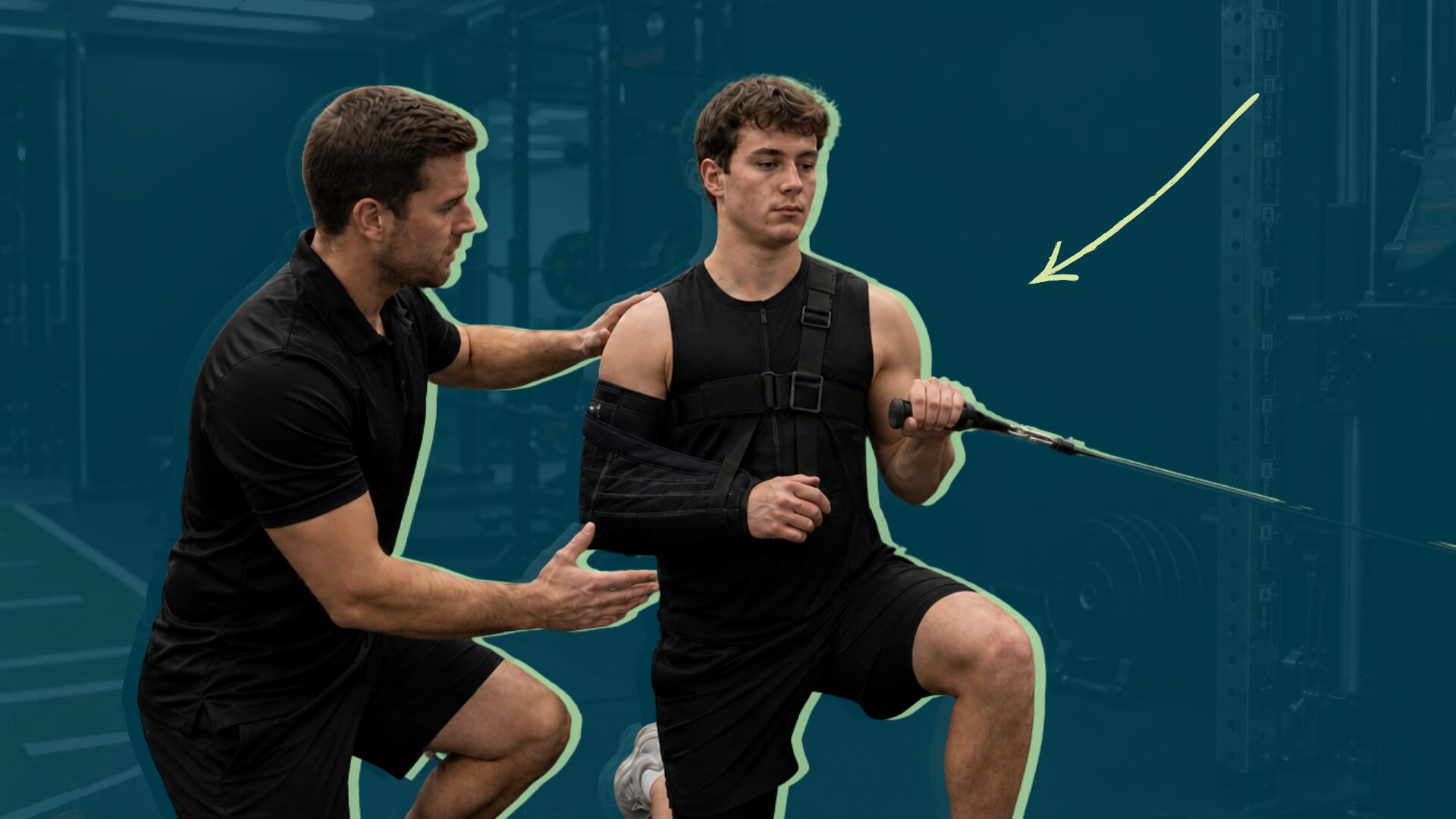
One of the most disruptive shifts in modern sports medicine has been abandoning absolute rest in favour of early mobility. Even in the management of acute soft-tissue injuries, the old RICE protocol has become completely obsolete. In its place: PEACE & LOVE, where the “L” stands for “load.” Recovery demands a rapid transition toward optimal loading, always guided by patient education.[3]
Keeping a patient immobile after orthopedic surgery doesn’t protect the structure that has been operated on. Quite the opposite. Immobilisation favours the emergence of persistent deficits that can compromise recovery and turn an acute dysfunction chronic.
The body’s response to surgery is as complex as it is aggressive. The body responds to the stress of anaesthesia, fasting, and the tissue injury itself through a cascade of endocrine, metabolic, and immunological changes. From a purely functional perspective, the first few post-operative weeks are usually accompanied by a predictable clinical picture: pain, inflammation, restricted range of motion, and muscle inhibition.
In our day-to-day clinical practice, the first step is to change how we interpret these symptoms. We shouldn’t see them simply as inevitable collateral damage from the trauma of surgery itself. They are the protective responses of a complex dynamic system. A “system of systems”, if you will.
Success in early rehabilitation doesn’t lie in prolonging rest, but in knowing how to actively modulate these variables from the very beginning.
Tweet ThisKeeping a patient immobile after orthopedic surgery doesn’t protect the structure that has been operated on. Quite the opposite. Immobilisation favours the emergence of persistent deficits that can compromise recovery.
@julio_caballe and Victor Jimenez Aransay
Cell biology is at the base of this approach through mechanotransduction. This is the process by which cells of the musculoskeletal system (such as osteoblasts and fibroblasts) need physical stimuli and mechanical loading to activate biochemical signals. Without that controlled mechanical deformation, it’s impossible to achieve optimal extracellular matrix synthesis or correct alignment of new collagen fibres.[1]
Enhanced recovery after surgery (ERAS) protocols demonstrate that immediate physical reactivation drastically reduces surgical stress and accelerates hospital discharge.[2]
When our patients walk into the clinic and ask us fearfully: “When will I be able to move?”, we always point out that the question is poorly framed. The real question we should be asking every morning is: what is the optimal load dose that this specific tissue can tolerate today without exceeding its biological capacity?
Road map to reactivation
To answer that question, we need to structure the patient’s path by dividing loading into four very clear biological phases based on physiological goals.
First is the immediate acute phase, which is the first 24 hours. Rehabilitation begins in the recovery room itself.
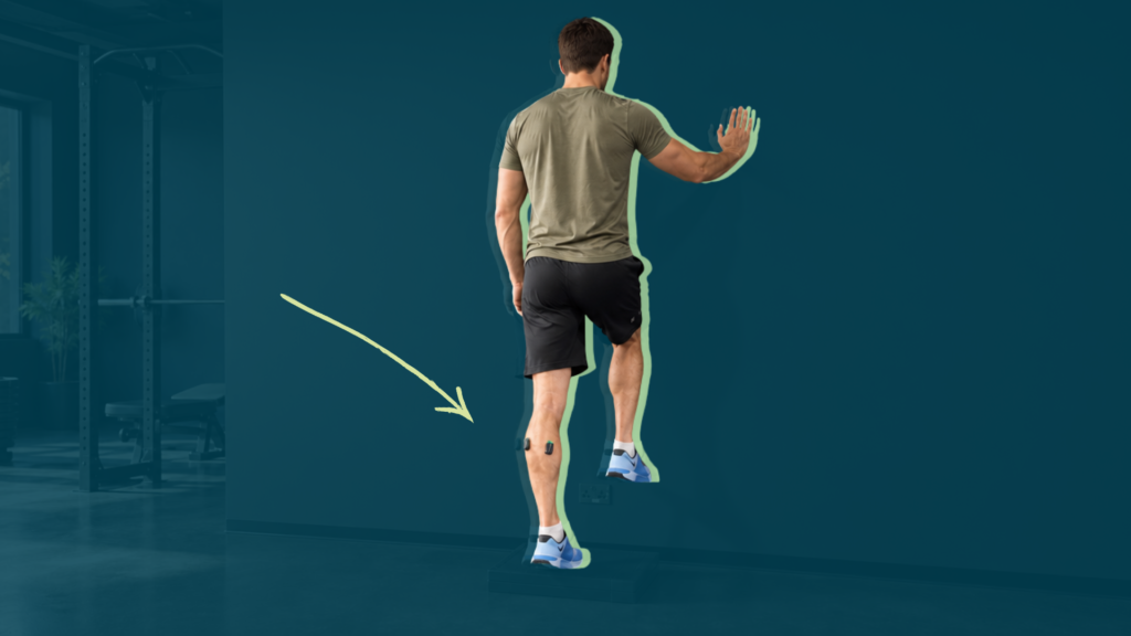
Between 4–6 hours after the effects of anaesthesia wear off, the top priority is to break bed immobility by encouraging early sitting and beginning weight-bearing as tolerated. With this early stimulus, we achieve a significant improvement in the systemic response to surgical stress, prevent disuse atrophy, and minimise the risk of serious vascular complications such as deep vein thrombosis.
The subacute hospital phase is from days 1–3. With the patient now on the ward, the focus shifts to the joint. We implement localised cryotherapy protocols to modulate pain and reduce inflammation, combined with independent walking around the hospital and flexion-extension mobilisations in bed (such as hip and knee flexion) to reduce pain. The critical objective at this stage is to prevent arthrogenic muscle inhibition (AMI).
Tweet ThisBetween 4–6 hours after the effects of anaesthesia wear off, the top priority is to break bed immobility by encouraging early sitting and beginning weight-bearing as tolerated.
@julio_caballe and Victor Jimenez Aransay
The initial remodelling phase spans the first four weeks. Once the patient is discharged and arrives at our physiotherapy clinic, mechanical loading must become progressive and directed.
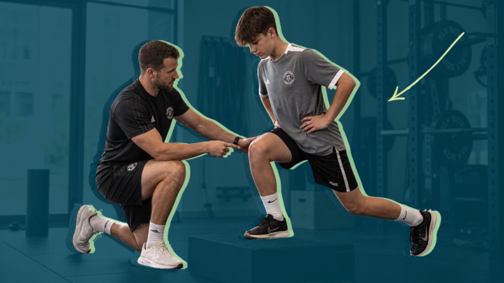
This is the time to re-educate the gait pattern and prescribe closed kinetic chain therapeutic exercises such as mini-squats or glute bridges. Supplement these with accessory cardiovascular work such as stationary cycling.
In this phase, mechanical stress stimulates cells to ensure, for example, correct bone fixation in the case of prostheses.
The functional re-adaptation phase lasts from week 4 onwards. After the first month, the tissue can tolerate greater tension. Treatment shifts toward strength training, progressively increasing the volume and intensity of loads, motor control work, dynamic stabilisation, and stationary cycling. The goal is to fully restore peripheral neuromuscular function.
Keeping muscles “switched on” post-surgery
The inability to properly activate muscles is one of the most frustrating situations in clinical practice, for both physiotherapists and the patient themselves.
Consider a scenario where, after a supraspinatus repair, we ask the patient to try to contract the deltoid and no visible contraction occurs. The patient’s immediate reaction is usually panic, thinking they’ve lost all their strength because of the surgery itself, or that the surgery has failed. However, the problem isn’t structural, but purely neurophysiological, as a protective mechanism of the body.
The trauma of surgery, combined with local inflammation, oedema, and post-operative pain, alters the afferent signals travelling to the central nervous system. This distortion generates reflex inhibition of alpha motor neurons. This phenomenon is AMI. AMI temporarily blocks the patient’s ability to voluntarily recruit muscle fibres, even though the muscle remains structurally intact.
The real danger of AMI is its persistence. If we don’t intervene immediately, AMI perpetuates weakness, makes the contraction deficit chronic, and, in the long term, silently accelerates the degeneration of joint cartilage.
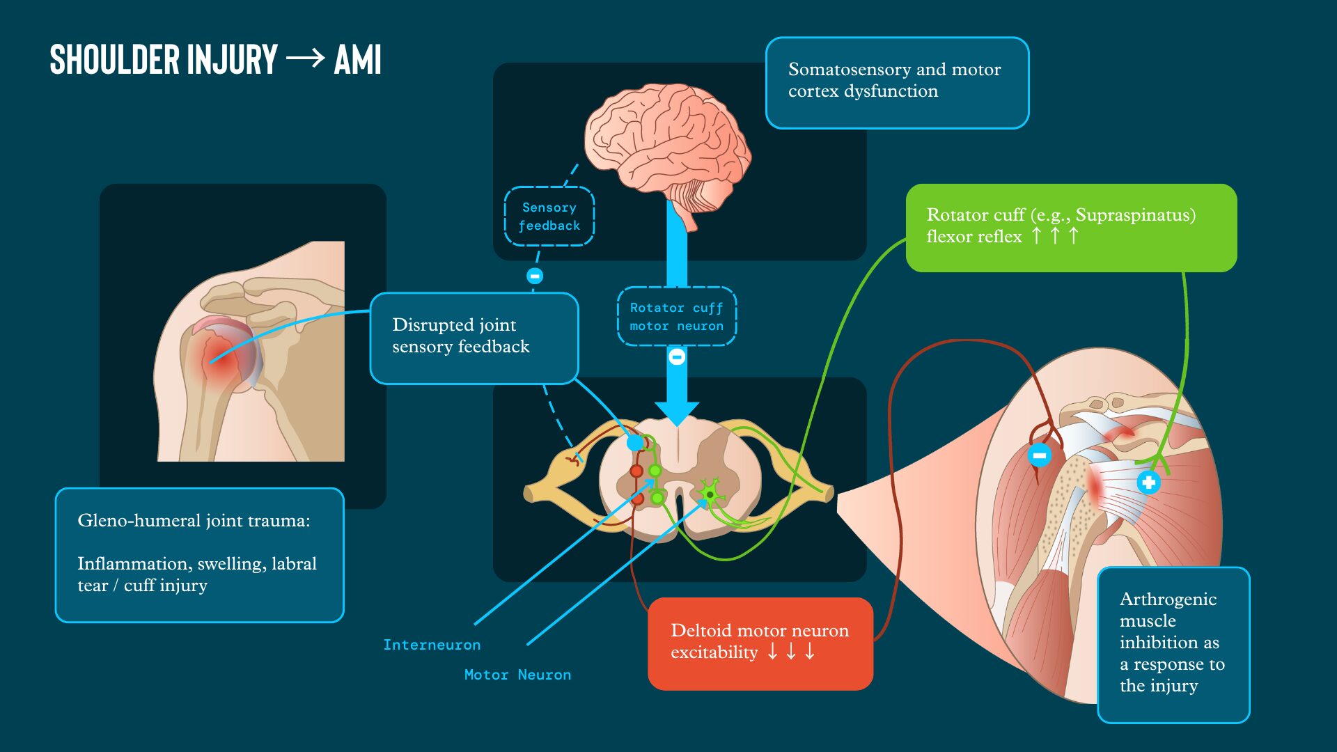
This often happens after surgical procedures. To resolve this block, we cannot make the mistake of pushing the patient through endless isometric contractions. We shouldn’t strengthen a muscle whose neural access is completely closed off. First, we need to “trick” the peripheral and central nervous system into reopening that communication pathway. We must hack the afferent signals reaching the spinal cord to suppress central inhibition before demanding voluntary effort.[4]
In our clinical practice, we approach AMI through two complementary neurophysiological pathways.
Peripheral modulation through cryotherapy entails applying local ice for 15 minutes right before starting the session. This drastically reduces nerve conduction velocity in joint receptors. It temporarily numbs the pressure and stretch signals causing the block, achieving an analgesic effect and reducing reflex inhibition at the spinal level.
Central modulation through cross-education involves training the healthy limb at very high intensities. This contralateral strength work triggers neural adaptations at the level of the motor cortex that travel downstream, attenuating inhibition and measurably preserving voluntary activation levels in the operated limb.
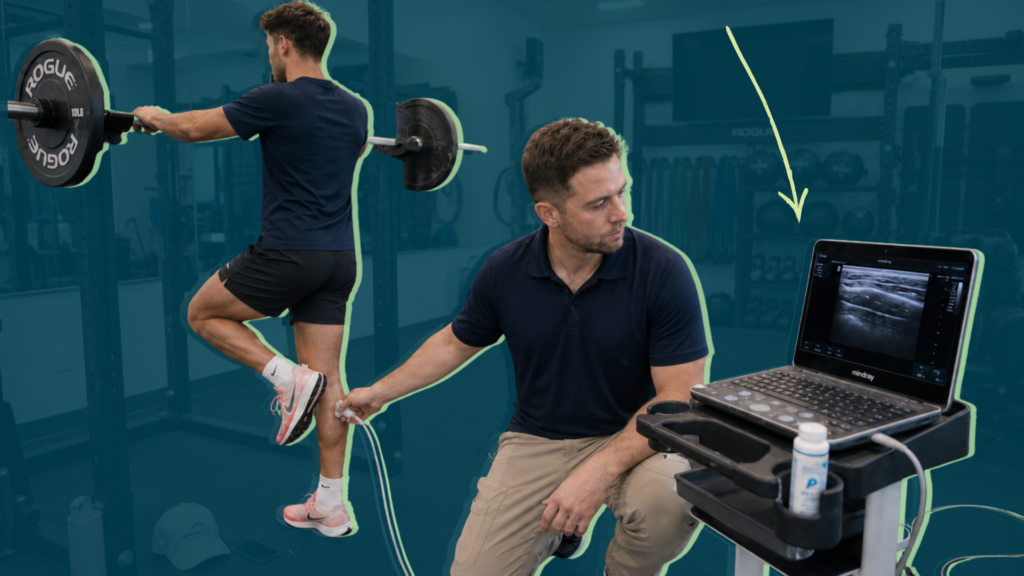
Beyond these two strategies, the day-to-day work relies on several modalities (Figure 2).

Neuromuscular electrical stimulation (NMES) is essential for triggering involuntary contractions and forcing recruitment of high threshold motor units when the brain is unable to send the command on its own.
Biofeedback is a valuable tool for cognitive re-education. By giving the patient a real-time visual signal of their minimal muscle electrical activity, we help them reconnect with the tissue and sharpen internal focus during contraction.
Virtual reality (VR) and motor imagery are “top-down” interventions, from the brain to the periphery. Working with directed mental visualisation of movement, or placing the patient in virtual environments, reactivates motor maps in the brain’s cortex, keeping those neural highways active even before the joint can physically move.
Low-load exercise and motor control progressively mobilise the joint without pain. These prioritise movement quality and precision of execution over resistance, re-teaching the nervous system to coordinate muscle co-activation without triggering pain alarms.
Tweet ThisPeripheral modulation entails applying local ice for 15 minutes before starting the session. This drastically reduces nerve conduction velocity in joint receptors achieving an analgesic effect and reducing reflex inhibition at the spinal level.
@julio_caballe and Victor Jimenez Aransay
Near-immediate movement allows for early loading
In orthopaedic rehabilitation, the answer to when to start loading tissue is always the same: as early as possible. Obviously, this must always remain within the limits of structural safety and the biological tolerance of the operated tissue.
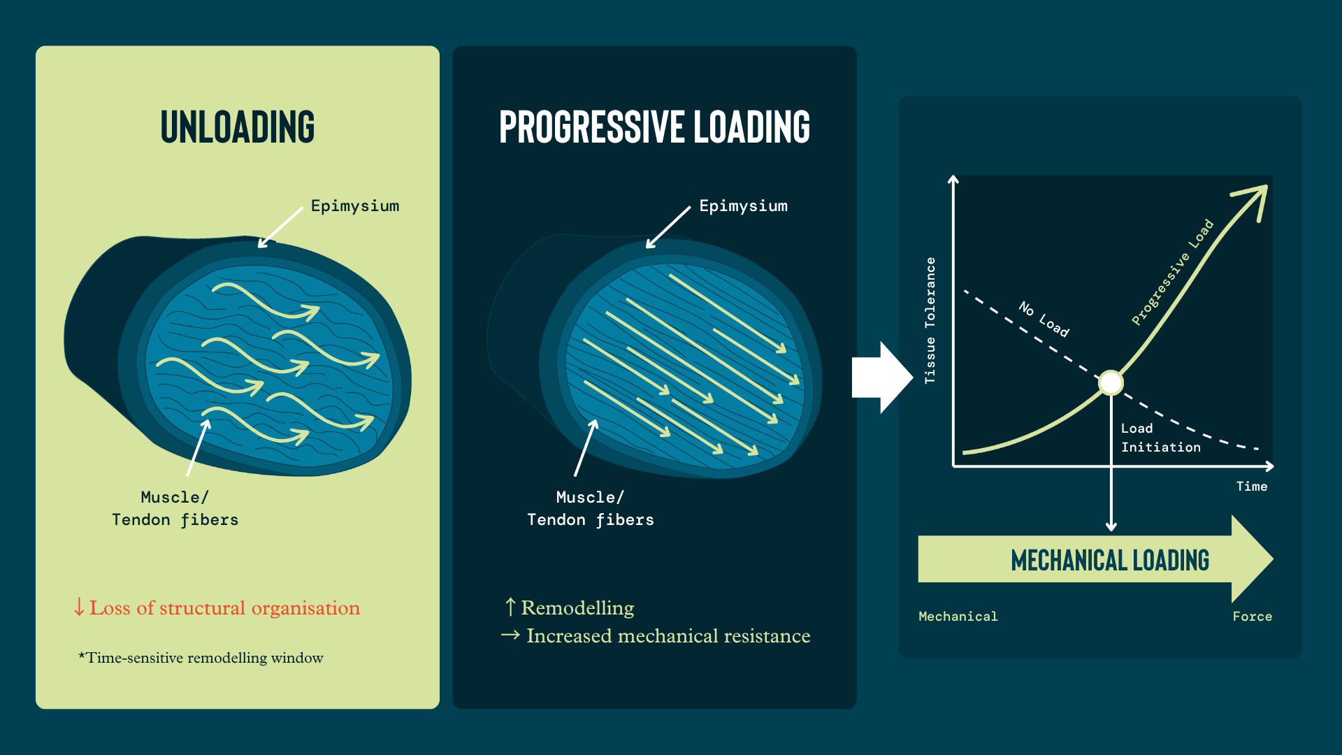
The timing of load initiation is a fundamental consideration. Musculoskeletal tissue is highly sensitive to mechanical stimulus. If we deprive a tendon, bone, or muscle of tension, an immediate loss of internal structural organisation occurs. Conversely, progressive loading is the biological trigger for its remodelling, increasing its mechanical resistance to future stress.[1]
In our clinic, we encounter a clinical pattern on a daily basis that significantly increases recovery timelines: fear of weight-bearing.
Watching a patient spend two weeks walking without bearing weight is, from a biological standpoint, a silent catastrophe. Just 5–14 days of complete unloading is enough to cause significant strength loss.[5,6]
However, the impact goes beyond losing centimetres of thigh circumference. The real disaster occurs in the nervous system if we choose complete unloading over early optimal loading.
Complete unloading reduces proprioceptive afferent input. The brain stops receiving the basic sensory feedback provided by ground reaction forces and joint pressure. This completely disrupts the motor schema. The brain loses representation of that leg or arm, seriously deteriorating neuromuscular control.
Tweet ThisIf we deprive a tendon, bone, or muscle of tension, an immediate loss of internal structural organisation occurs. Conversely, progressive loading is the biological trigger for its remodelling.
@julio_caballe and Victor Jimenez Aransay
Compensatory and protective patterns develop as a result, making AMI chronic and perpetuating issues that become much harder to resolve later on.
Early optimal weight-bearing loading allows us to preserve functional motor maps in the brain. By continuously stimulating the somatosensory and motor cortex, we avoid that negative cortical reorganisation induced by disuse. Peripherally, this maintains healthy intermuscular coordination, preserving co-activation patterns and the reaction times of the joint’s dynamic stabilisers.
Our mission in the early phases isn’t to avoid loading so that “nothing happens.” On the contrary, we actively seek the optimal mechanical stimulus to maintain system balance without putting the surgery at risk.
Using symptoms as markers of progress
Monitoring real progress requires looking beyond the treatment table. Progress should be measured mainly through the symptom response to loading, supported by clinical observation.
Positive progress is when pain decreases, functional capacity and range of motion increase, and the patient shows progressive tolerance to daily activity — for example, by increasing step count.
The criteria for progressing load are stable or decreasing pain during and 24 hours after mechanical exposure; absence of joint swelling flare-ups; and sustained movement quality and motor control, i.e., no mechanical compensations or limping.
Conversely, the criteria for stopping or regressing load are an increase in pain above 3/10 on the visual analogue scale that lasts more than 24 hours; onset of morning joint stiffness or visible inflammation after the session or walking; and the inability to maintain voluntary muscle activation during the task (recurrence of AMI).
At this point in post-surgical recovery, the common danger is the opposite of what it was before: many patients tend to overestimate their healing as soon as symptoms decrease.
A classic scenario we often see in the clinic is patients getting up pain-free, growing overconfident, and multiplying their activity: taking a long walk after knee surgery, or starting to lift weights at home after a shoulder repair.
The next day they come back to the clinic with a pain flare-up, exacerbated inflammation, severe stiffness, and functional limitation much worse than in previous days. This happens because the tissue’s biological capacity hasn’t yet structurally adapted to that tension spike, no matter how much symptoms had temporarily improved.
Because of this tendency and its consequences, we need to educate the patient to understand that progression in physiotherapy must be guided by quantified, controlled exposure to load, never by how they feel on a single “good day.”
Tweet ThisThe criteria for progressing load are stable or decreasing pain during and 24 hours after mechanical exposure; absence of joint swelling flare-ups; and sustained movement quality and motor control, i.e., no mechanical compensations or limping.
@julio_caballe and Victor Jimenez Aransay
Strategies for the pre- and post-operative period
High-quality rehabilitation in the acute phase isn’t limited to prescribing exercise. There are systemic behavioural and metabolic factors that tip the balance toward clinical success or failure. Sleep, nutrition, stress regulation, and sunlight exposure are critical modulators of pain, immune function, inflammation, and the speed of tissue repair. These are often undervalued variables but optimising them makes a decisive difference.
Sleep is, without doubt, the most powerful biohacking tool. The scientific evidence is overwhelming: poor sleep quality not only suppresses the immune system, but amplifies pain perception and delays biological healing processes.[7]
Likewise, the post-surgical metabolic environment demands precise nutritional support. Optimised protein intake and an adequate energy balance are essential pillars to slow the loss of muscle mass caused by the inactivity period and to speed up soft tissue healing.
Under this same systemic perspective, we must understand that true recovery doesn’t start after surgery, but weeks before entering the operating theatre. The goal of pre-habilitation is to optimise the patient in three critical areas so their body can respond reliably to the enormous surgical stress and achieve superior post-operative function.
First is their preoperative physical condition. Proactively seeking to improve strength, mobility, and neuromuscular control before the procedure is essential. Entering the operating theatre with stronger, more functional musculature (such as a powerful quadriceps before knee arthroplasty) is the number one predictor of faster recovery and greater independence in the early stages.[8]
Next is the metabolic environment and habits. Patients should maximise protein intake and ensure quality rest before the operation. At this stage, completely quitting smoking is non-negotiable: nicotine strangles tissue perfusion, severely impairs healing, worsens bone consolidation, and exponentially increases the risk of infections or post-operative complications.[9,10]
Third are the psychosocial factors and education. Managing expectations is essential. When we thoroughly explain to the patient what the surgical process will be like and what steps we’ll take in immediate rehabilitation, we drastically reduce their anxiety and defuse fear of movement (kinesiophobia).
An educated patient is a patient who doesn’t freeze up when faced with acute pain and who trusts the optimal loading process.[11]
Patients who approach surgery with optimised physical condition, healthy biological habits, and realistic, mature expectations experience dramatically better functional outcomes.
The key to success in early rehabilitation isn’t waiting for tissue to heal passively, but preparing the biological and neurophysiological ground so that movement can do its job from day one.
Tweet ThisSleep, nutrition, stress regulation, and sunlight exposure are critical modulators of pain, immune function, inflammation, and the speed of tissue repair. These are often undervalued variables but optimising them makes a decisive difference.
@julio_caballe and Victor Jimenez Aransay