
Anyone reading this likely knows at least one athlete who has suffered an ACL injury. Emerging scientific research and clinical experience are disrupting the idea that surgery is the essential step that connects injury to rehabilitation. Even so, ACL repair surgery is still the standard option.
Publication bias keeps this default setting alive. There is a significantly larger body of research focused on ACL surgery compared to conservative—that is, non-operative—management or the pre-operative phase.
The pre-operative phase should be where a multidisciplinary team and the athlete develop the evidence to make an informed decision regarding surgery, or whether their injury, return to play, and future athletic career can all be managed successfully without surgery. It can also function as “pre-surgery prehab,” setting the athlete up for better post-surgical outcomes.
Tweet ThisAnyone reading this likely knows at least one athlete who has suffered an ACL injury. Emerging scientific research and clinical experience are disrupting the idea that surgery is the essential step that connects injury to rehabilitation.
@FrouinAntoine
While the first high-quality studies on pre-operative management started to emerge around 10 years ago, robust evidence for conservative management may take another decade to fully develop.
However, clinical practice shouldn’t always wait for long term data when there are already well-identified cases where conservative treatment could be a valid alternative to surgery. The ACL SNNAP study [3] demonstrated that when conservative treatment is chosen instead of immediate surgery, patients achieve similar quality of life outcomes at one year post-injury.
This approach has the potential to improve return to performance outcomes, and make the most fundamental decisions in ACL rehab truly individualized.
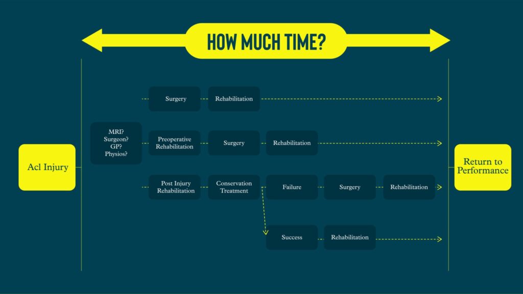
Post-ACL treatment pathways
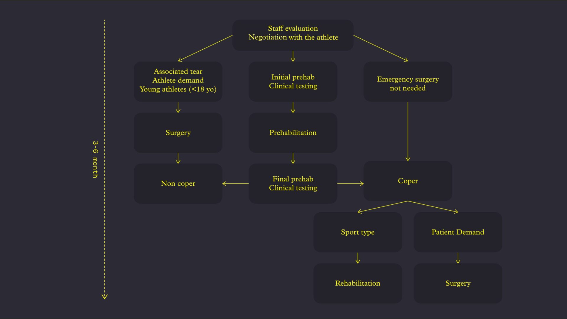
Before rushing into ACLR surgery, we should screen athletes to see if they are “copers”: suitable candidates for conservative treatment.
Stability is the overarching factor distinguishing between copers and non-copers. Non-copers are unlikely to recover sufficient knee stability, even when following the best rehabilitation, combined with optimal strength & conditioning programs, and sport-specific training.
Some studies already provide clinical criteria to help differentiate between copers and non-copers. Fitzgerald et al. [2] proposed a set of benchmarks. Copers will achieve at least 80% between-limb symmetry in the timed 6-meter hop test; 80% on the Knee Outcome Survey – Activities of Daily Living (KOS-ADL); 60% or higher on the Global Knee Rating Score; and report no more than one episode of giving way.
We can and should go further in evaluating athletes’ capacities, whether in a conservative management pathway or during the preoperative phase.
Moreover, managing an athlete with an ACL tear goes far beyond simply restoring strength, loading capacity, and sport-specific skills. It is a far more complex process. ACL rehabilitation is one of the most underestimated and challenging areas within musculoskeletal rehabilitation.
The long term consequences of surgery on muscle and tendon are often underestimated or overlooked. Even after extended recovery periods, tendons are not fully healed, and muscle volume is not completely restored. [8] Specifically, when the semitendinosus and gracilis tendons are harvested for grafts, only a minority of athletes experience true tendon regeneration. Many will never recover a distal insertion, meaning these muscles will no longer contribute effectively to joint function.
One clear consequence of this underestimation is the low rate of athletes who successfully return to their previous level of performance after ACL injury—only about 55%, which is among the lowest return to sport rates for any musculoskeletal injury. [10]
About 40% of clinicians do not use any formal return to play (RTP) criteria. [11] Among the others, 32% rely solely on time, 15% combine time with some basic criteria, and only 13% use objective measures such as strength, functional capacity, or psychological readiness.
This likely extends beyond clinical settings into strength & conditioning practices as well.
A proper test-retest approach should not only be applied at key milestones, but should be integrated continuously throughout the entire rehabilitation process. Regular assessments motivate athletes and practitioners, while also providing critical feedback to adjust goals and modify training if progress is not on track.
Pre-op evaluations
In the era of Instagram, TikTok, and content designed to be easily consumable, everything is made to look simple and straightforward. But anyone working with real athletes and real injuries knows that rehabilitation is anything but simple.
If you don’t assess the athlete’s specific impairments and needs, you end up providing generic exercises.
The assessments here focus solely on the prehabilitation phase. They provide us a clear understanding of each athlete’s specific issues, contributing data that will either provide a roadmap for post-operative rehab, or create the opportunity to guide the athlete toward a conservative treatment pathway, avoiding the outdated straight-to-surgery approach.
Athletes’ internal state and external outcomes
Patient-reported outcome measures (PROMs), such as standardized questionnaires, are often undervalued.
However, they provide critical insights into an athlete’s subjective experience and progression throughout rehabilitation. Their ease of administration and interpretation makes them indispensable in both clinical practice and research. Neglecting such questionnaires is a missed opportunity to comprehensively monitor recovery and guide individualized treatment plans.
The Hospital Anxiety and Depression Scale (HAD [16]) acknowledges the likelihood that athletes may experience symptoms of anxiety or depression following an ACL injury.
During the recovery period, the athlete is removed from the team environment and training routines, which drastically changes their personal and social context. Their primary concerns often narrow down to questions like, “How is my knee today?” and “When can I get back on the field?” Despite ACL injuries being one of the most common knee injuries in football, I am often surprised when athletes ask if they can start running just two weeks post-surgery.
Accepting that rehabilitation will be a long, demanding process is difficult. Moreover, in some cases where rehabilitation progress is minimal, underlying depression may be a contributing factor.
Tweet ThisAbout 40% of clinicians do not use any formal return to play criteria. Among the others, 32% rely solely on time, 15% combine time with some basic criteria, and only 13% use objective measures such as strength, functional capacity, or psychological readiness.
@FrouinAntoine
ACL rehabilitation is inherently multidisciplinary. That includes sport psychologists or mental coaches, as strength & conditioning coaches or physiotherapists alone cannot address all aspects of recovery.
The Knee Injury and Osteoarthritis Outcome Score (KOOS [17]) is divided into five subscales: symptoms, pain, activities of daily living, sports / recreation, and quality of life.

The questionnaire scores each individual domain and provides an overall score out of 100%. The KOOS helps highlight deficits or changes in the athlete’s perception of their knee that they may not verbally express during sessions. Quantifying these subjective feelings often reveals concerns that remain unspoken.
Finally, there is the Anterior Cruciate Ligament Return to Sport after Injury scale (ACL-RSI [18]). Although this tool is not validated for preoperative or conservative treatment phases, it is widely used to assess psychological readiness for return to sport post-surgery. Despite the lack of formal validation in earlier phases, it is valuable to gauge an athlete’s mental preparedness for key rehabilitation milestones, such as returning to jumping, sport-specific skills, sprinting, and changes of direction.
A KOOS and ACL-RSI score of at least 70% are benchmarks for return to running and jumping, 90% before progressing to sport-specific skills, and ideally 100% before full return to performance and competition, alongside good strength and functional recovery.
Tweet ThisIn the era of Instagram, TikTok, and content designed to be easily consumable, everything is made to look simple and straightforward. But anyone working with real athletes and real injuries knows that rehabilitation is anything but simple.
@FrouinAntoine
Putting a number on pain
Monitoring knee-related pain is just as critical as tracking any other rehabilitation parameter. However, it is often underestimated or neglected by clinicians and performance staff. Relying on subjective impressions rather than objective measurements is a common mistake. A number is always more reliable than a guess.
Systematically assessing pain before and after each session provides valuable insights into both the mechanical load applied to the knee and the athlete’s perception of that load. This information allows for precise adjustments to the rehabilitation process.
An important point to emphasize is that pain progression should be interpreted in context. If an athlete’s physical capacities are improving—through increased mechanical loading or more demanding tasks—and pain levels remain stable or do not increase, this represents progress. The knee is demonstrating improved tolerance to stress without amplifying nociceptive signals.
Additionally, it is essential to consider that pain fluctuations may sometimes result from the discontinuation of pain management strategies, such as stopping anti-inflammatory medications or analgesics. Increases in pain under these circumstances are not necessarily indicative of mechanical overload or tissue irritation, but may reflect the withdrawal of pharmacological support.
Tweet ThisACL rehabilitation is inherently multidisciplinary. That includes sport psychologists or mental coaches, as strength & conditioning coaches or physiotherapists alone cannot address all aspects of recovery.
@FrouinAntoine
Knee effusion
Alongside pain monitoring, knee effusion remains one of the most critical parameters in the early post-injury phase. A primary objective during this period is to achieve and maintain a “dry knee,” as persistent effusion is often associated with delayed recovery, reduced neuromuscular control, and impaired progression through rehabilitation milestones.
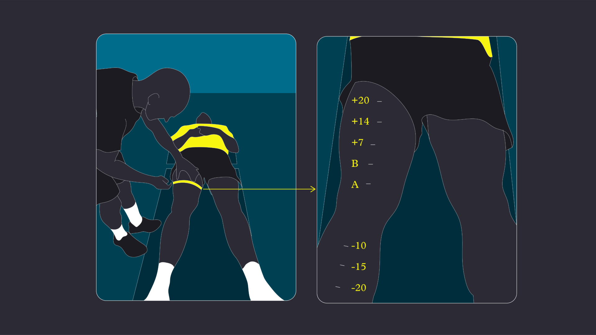
Effusion is typically assessed through circumferential measurements above and below the patella, using well-defined anatomical landmarks that offer reliable and reproducible data in clinical practice. Specific clinical tests such as the stroke test can confirm the presence or absence of joint effusion with good sensitivity. [19]
Systematically monitoring effusion provides crucial feedback on the knee’s response to mechanical loading and helps guide the progression or regression of rehabilitation exercises when necessary.
If the knee is not dry, there is an ongoing risk, not only in terms of joint health but also due to effusion’s direct impact on neuromuscular function.
Persistent effusion contributes to arthrogenic muscle inhibition (AMI), which significantly reduces the capacity for muscle activation and force production. AMI is not merely a local phenomenon at the knee but involves central mechanisms—including at both spinal and supraspinal levels. [21] This makes it critical to systematically assess and reassess effusion, particularly how it evolves in response to physiotherapy interventions and training loads.
To help athletes conceptualize this, I often use the analogy of a fluorescent light tube filled with neon to represent the muscle. Some of the neon lights (motor units) are no longer functioning, and the electrical current—supplied by the nervous system—is disrupted. This faulty supply affects the number of lights that can be switched on, resulting in reduced brightness, representing diminished muscle contraction and force production.
Tweet ThisIncreases in pain under these circumstances are not necessarily indicative of mechanical overload or tissue irritation, but may reflect the withdrawal of pharmacological support.
@FrouinAntoine
The goal of rehabilitation is not only to restore muscle function (neuromuscular control) but also to recover muscle volume. Muscle atrophy begins almost immediately after injury due to reduced activation. Regularly and systematically assessing muscle volume loss throughout rehabilitation is essential. It provides valuable insight into both the structural and functional consequences of the injury.
While the direct causal link between AMI and muscle atrophy has not yet been fully established, current evidence suggests that muscle volume is the visible part of the iceberg. The underlying neuromuscular dysfunction is the submerged part.
Muscle volume
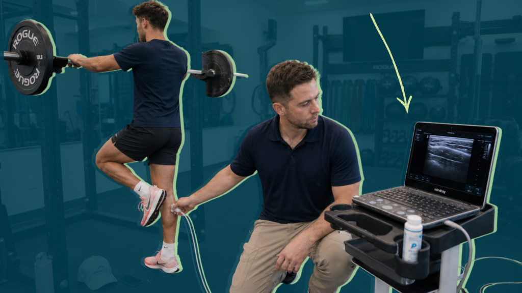
3D ultrasound has emerged as a promising alternative to MRI, offering greater accessibility and lower costs, while being reliable for measuring thigh and calf muscle volumes. [24, 25] However, 3D ultrasound is time consuming and not cost effective for routine clinical use. However, advancements in artificial intelligence may accelerate this process. [27] This creates the potential for regular monitoring of muscle volume to be more accessible and efficient during rehabilitation, particularly for athletes recovering from ACL injuries.
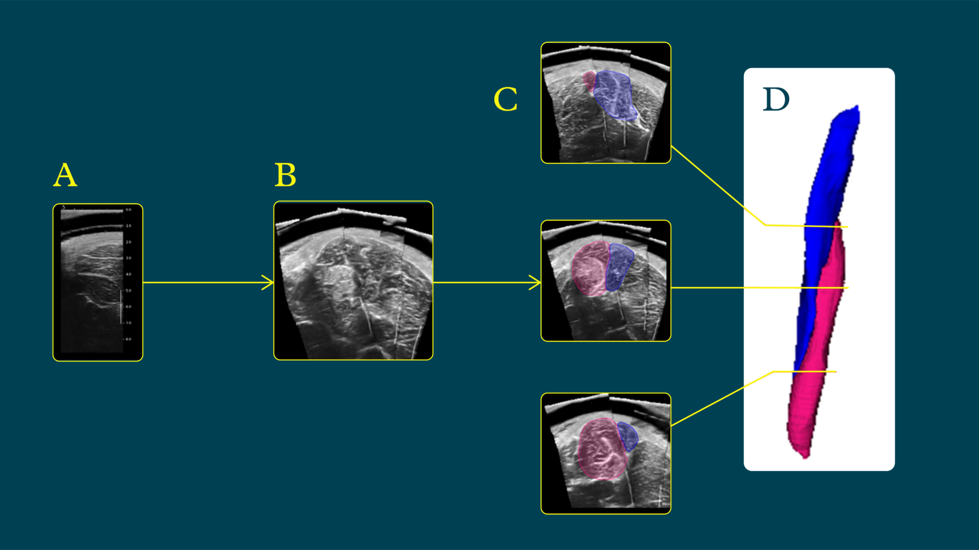
The most relevant muscle groups to monitor during ACL rehabilitation are the thigh and calf muscles, given their critical roles in knee stability and performance.
In clinical settings, circumference measurements are a simple yet valuable tool. For the thigh, measurements are at 7, 14, and 20 cm above the superior border of the patella, targeting both quadriceps and hamstrings. For the calf, measurements are performed 10, 15, and 20 cm below the tibial tuberosity.
While this method is prone to certain biases and limitations, it remains a more objective assessment than purely visual estimations. However, one of the main limitations of circumference measurement is that it does not differentiate between muscle mass, adipose tissue, or edema.
To address this, pair the circumference assessment with skinfold caliper measurements, specifically to monitor adipose tissue accumulation on the injured limb. This helps determine whether apparent size preservation may actually reflect increased fat rather than muscle retention.
The three relevant skinfold sites are mid-thigh lateral, at 50% of the femur length, lateral aspect; vastus medialis oblique, at 45° relative to the mediolateral axis of the patella; and posteromedial thigh, targeting the adductor and semimembranosus region, approximately 10 cm below the gluteal fold.
The objective is to monitor changes in thigh composition throughout the rehabilitation process. Muscle atrophy, combined with a reduction in physical activity, could be accompanied by an increase in subcutaneous fat in the affected limb (osteoarthritis or knee pain).[28] This approach allows for a more comprehensive evaluation of muscle recovery beyond simple volume metrics, providing insight into both morphological and compositional changes.

Range of motion
All major consensus statements and clinical guidelines regarding ACL rehabilitation emphasize that full recovery of knee range of motion (ROM), particularly full extension, is a non-negotiable prerequisite for successful rehabilitation and return to sport. [10,12,13,29] Despite this, I still observe athletes with a persistent knee extension deficit of 5-10°, even during the preoperative phase. Failure to restore full extension can lead to significant downstream consequences if not addressed pre-operatively: altered gait mechanics, quadriceps inhibition, increased joint loading, and potentially compromised surgical outcomes. This highlights the critical importance of systematically and frequently assessing ROM, including in comparison to the contralateral limb, from the earliest stages of pre-op rehabilitation through the post-operative stage.
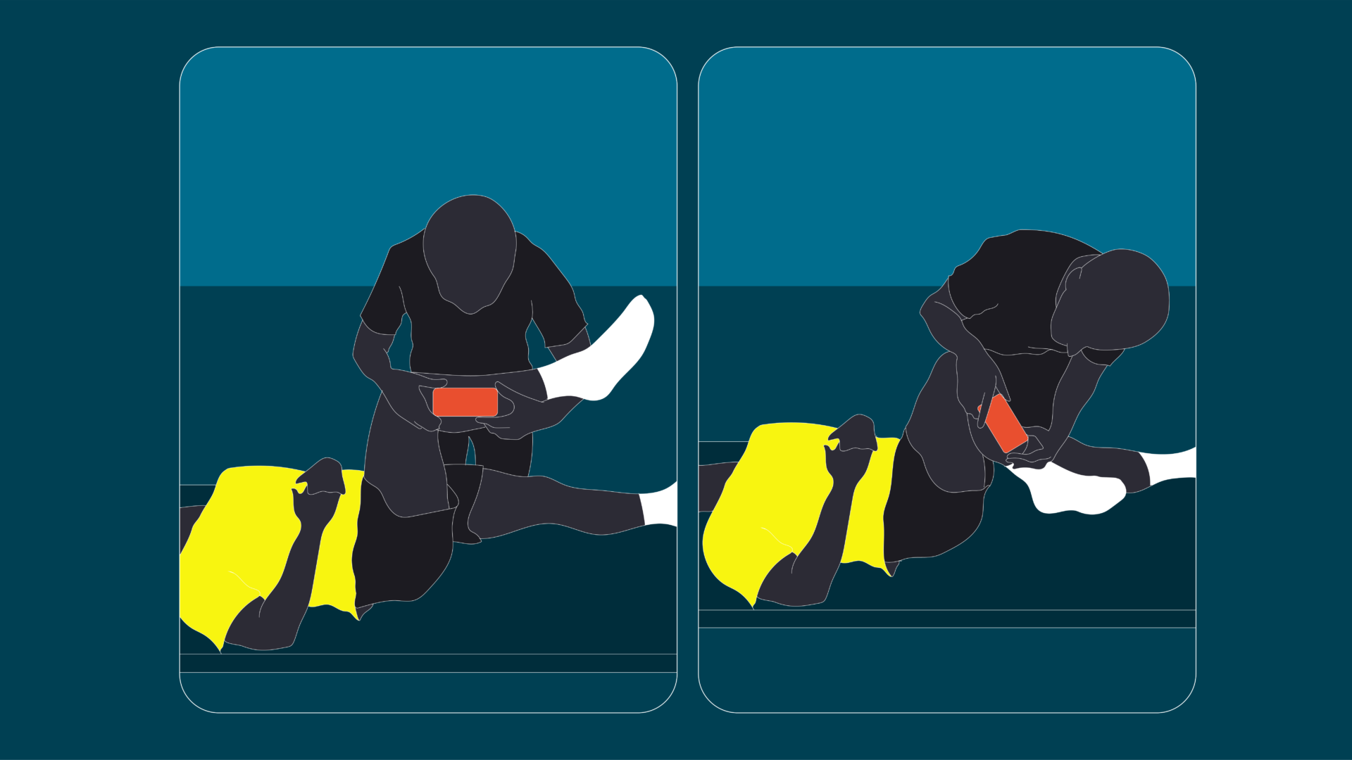
While knee flexion and extension are routinely measured in clinical practice, internal and external tibial rotation should also be systematically assessed.
Tibial rotation plays a fundamental role in knee joint function, particularly in tasks involving high mechanical demands, such as deceleration, pivoting, and rapid changes of direction.
Impairments in tibial rotation—whether due to capsular stiffness, soft tissue restrictions, or neuromuscular dysfunction—can significantly alter knee biomechanics.
Specifically, restrictions in tibial rotation may lead to increased joint reaction forces, greater strain on the passive structures of the knee, and elevated risk for compensatory movement patterns. This is particularly relevant during high speed decelerations, where proper tibiofemoral alignment is crucial to dissipate forces safely.
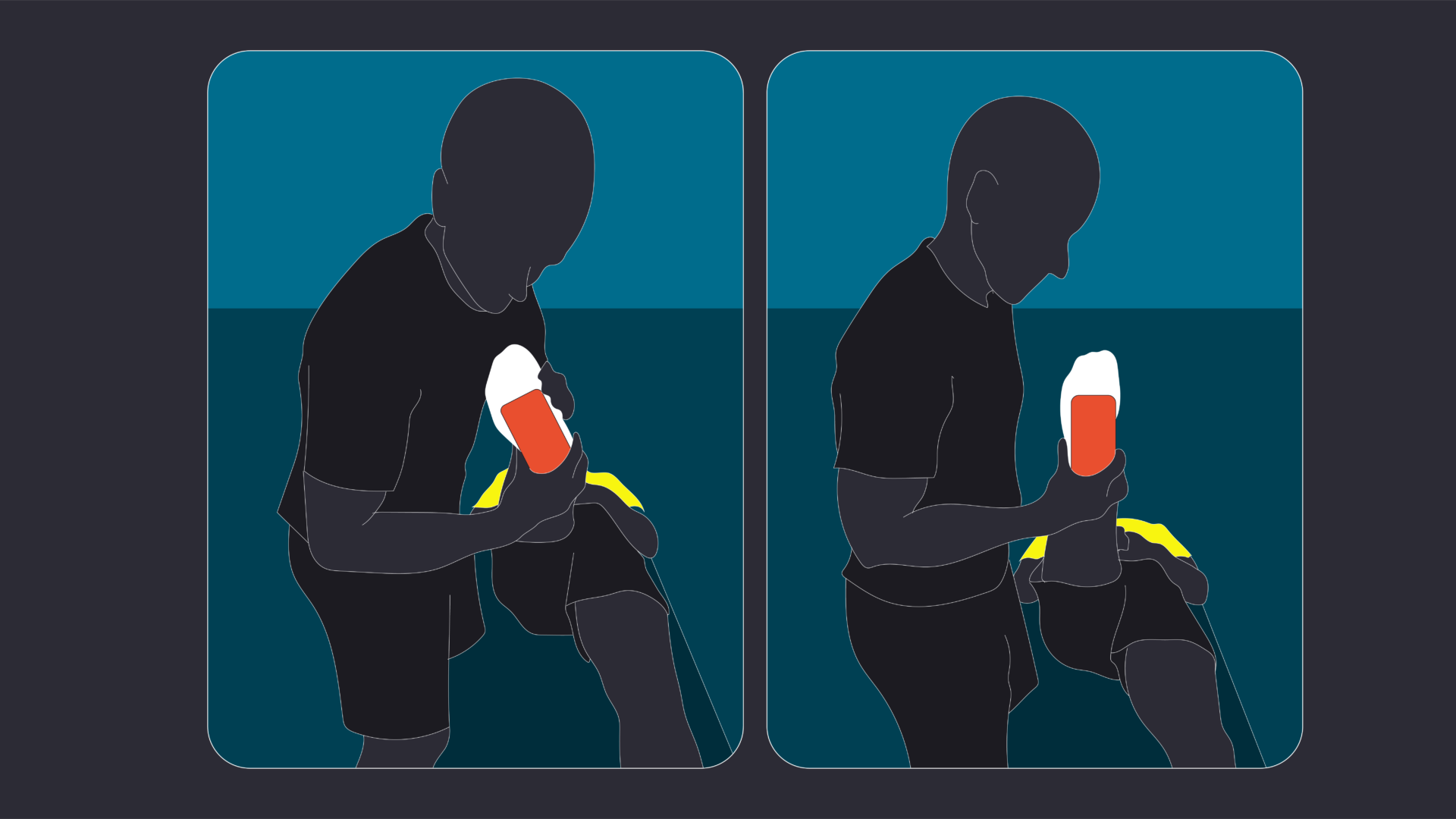
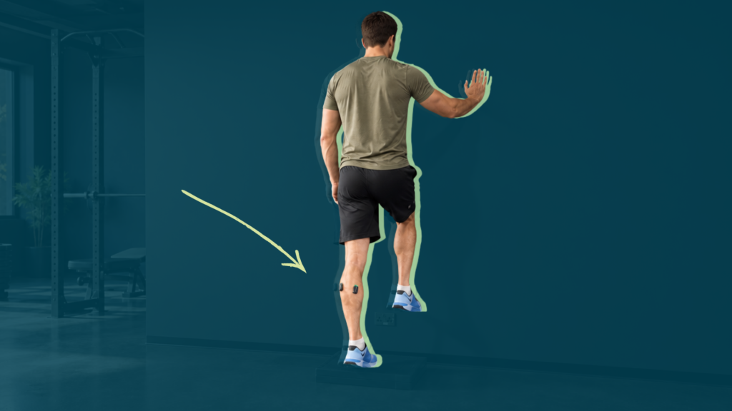
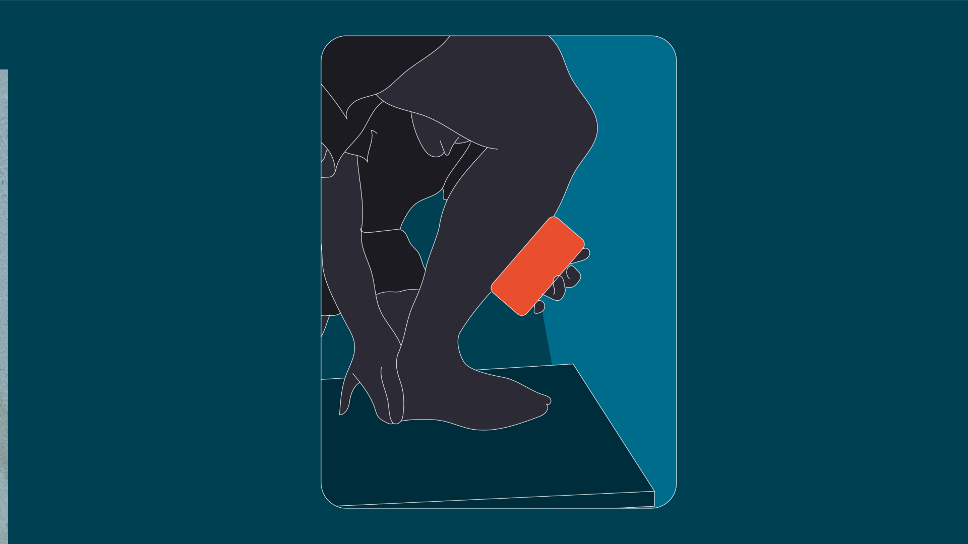
Adequate dorsiflexion is strongly correlated with proper lower limb biomechanics, particularly during tasks involving landing, deceleration, and changes of direction. Therefore, the final range of motion assessment is ankle dorsiflexion, via the Weight Bearing Lunge Test (WBLT). [30] This test provides a reliable measurement, either in degrees or in centimeters, based on the distance from the big toe to the wall (see Figure 9).
Normative values generally range from 9-12 cm in the general population. [31]
Ask the athlete to bear their full weight on the tested limb while moving the knee forward towards the wall, aiming to touch it without lifting the heel. A valid attempt requires the calcaneus to remain in full contact with the ground throughout the movement.
Any suspicion of hip mobility restrictions should be assessed during the preoperative phase, as well. However, I tend to prioritize this evaluation in the postoperative phase unless I observe a clear limitation earlier.
Tweet ThisMoreover, managing an athlete with an ACL tear goes far beyond simply restoring strength, loading capacity, and sport-specific skills. It is a far more complex process. ACL rehabilitation is one of the most underestimated and challenging areas within musculoskeletal rehabilitation.
@FrouinAntoine
Assessing muscle function
After an ACL injury, whether during the prehabilitation phase prior to surgery or when considering a conservative treatment approach, these assessments are the foundation for an individualized rehabilitation program.
Muscle activation
There is an ongoing debate about using electromyography (EMG) in clinical settings. I use EMG exclusively as a biofeedback tool, rather than as an objective evaluation method. This is primarily because the EMG systems typically available in clinical practice are not comparable to those used in research laboratories in terms of signal quality and reliability.
Additionally, the necessary preparation protocols for reliable EMG acquisition—such as skin shaving, cleaning with alcohol, and precise electrode placement following SENIAM recommendations—are often not fully implemented in clinical contexts. Without adhering to these standards, the validity and consistency of the EMG signal are compromised.
Moreover, a critical step often overlooked in clinical settings is the normalization of EMG signals to the maximum voluntary contraction (MVC) or maximum EMG output of each muscle. This is essential to allow for meaningful comparisons within and between individuals.
It is also important to note that the relationship between muscle activation levels and hypertrophy outcomes remains unclear. [33,34]
While EMG can be a useful tool for real time feedback in rehabilitation or strength training, it should not be considered a reliable evaluation or predictive tool at this stage, especially in clinical environments.
Iso strength measurements
The isokinetic dynamometer is considered the gold standard for strength assessment. Evaluations are ideally conducted at two concentric angular velocities, typically 60°/s and 240°/s, and one eccentric velocity (30°/s), allowing comprehensive analysis of the torque-angle relationship.
Our protocol prioritizes testing at the slower concentric (60°/s) and eccentric (30°/s) velocities, as they provide greater reliability in assessing maximal voluntary strength. Insufficient baseline strength compromises the development of subsequent neuromuscular qualities. Therefore, our primary objective is to establish maximal strength and slow concentric strength before progressing to speed-strength, power, and explosiveness.
A critical methodological consideration is comparing strength at consistent joint angles. While some clinicians emphasize peak torque values, comparing peak torques obtained at different joint angles is misleading. The preferred approach involves angle-specific torque comparisons to accurately quantify deficits. [37]
Notably, recent evidence supports the reliability of hand-held dynamometry for strength assessment post-ACL injury or reconstruction. Normative strength values range from 6-7 times bodyweight for knee extension and 3–4 times bodyweight for knee flexion. [38] Given its reduced time demands, HHD may be advantageous for frequent re-evaluations.
For strength testing, the standard knee extension angles are 80°, 45°, and 20°; for knee flexion, 30°, 60°, and 110°. Including measurements at shorter muscle lengths (e.g., 20° extension and 110° flexion) is essential, as deficits may persist in these positions despite apparent recovery at mid-range angles.
To ensure validity, the dynamometer must be securely fixed to minimize tester-induced artifacts, and the hip must be stabilized to prevent compensatory movements. Preceding the assessment is a standardized warm-up and familiarization protocol consisting of submaximal contractions at 50%, 70%, and 90% of perceived maximal effort, followed by three maximal voluntary contractions at each angle. Rest intervals of 1.5-2 minutes between trials ensure adequate neuromuscular recovery.
Given that HHD measures unidirectional force, resistance should always be applied perpendicular to the limb segment to capture the total force output accurately.
To mitigate the influence of the stretch-shortening cycle and tendon elastic recoil, a pre-tension corresponding to approximately 10% of the maximal voluntary contraction of the contralateral limb is applied prior to maximal testing.

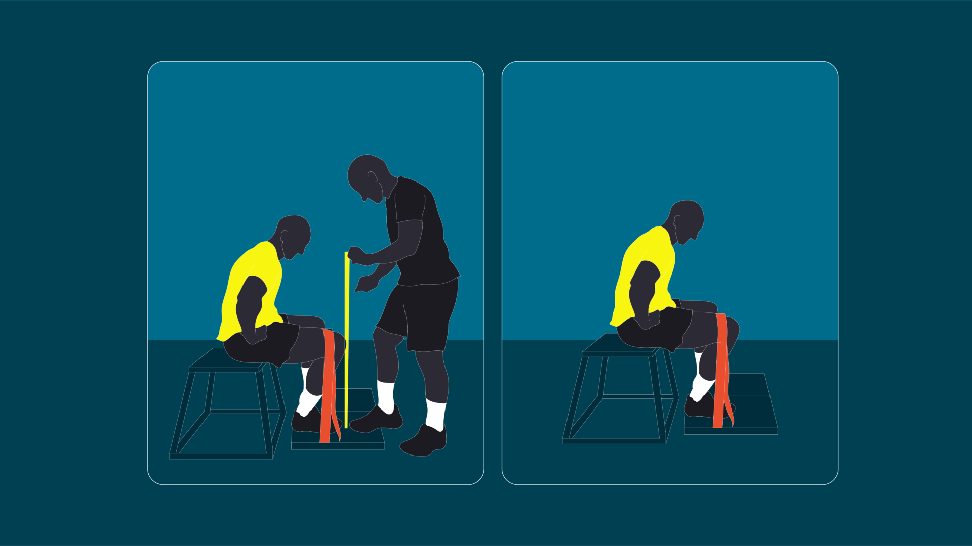
Two additional measurements are plantar flexion strength (soleus) and calf strength endurance (heel rise test [39]), as these muscles contribute significantly to reducing ground reaction forces and anterior tibial translation. [40]
Assess isometric soleus strength with the foot dorsiflexed. Vertical alignment between knee and foot can help standardize dorsiflexion between limbs, but there is potential measurement bias related to this positioning. Normative values for soleus strength are approximately 1.8 times bodyweight. [41]
Dynamic evaluations: Deceleration, jumping, and landing
These assessments are the primary evaluations applicable to nearly all athletes. Once the knee is free of both pain and effusion, dynamic evaluations can begin.
The objective at this stage is to prepare the athlete for propulsion and shock absorption tasks, with an initial focus on their capacity to decelerate effectively.
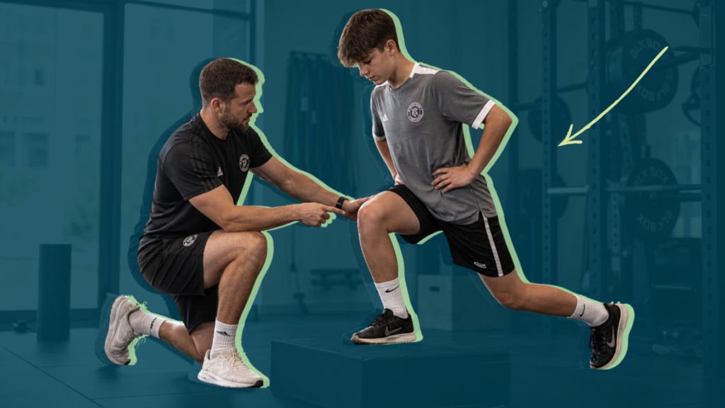
The progression typically begins with static standing assessments, followed by bilateral and unilateral squats. Provided that symmetry and balance between limbs are satisfactory in these tests and the athlete shows adequate readiness and physical condition, more demanding dynamic evaluations, such as the countermovement jump and drop jump, can be incorporated.
We developed the Knee Absorption Test to fill in the gap for specifically assessing deceleration capacity following balance evaluation during a bodyweight squat. This test involves performing a high velocity squat with an active braking phase occurring near 90° of knee flexion.
Both intraday and interday reliability of the KAT are excellent. The braking phase parameters, such as peak force and braking power, can be readily quantified without exposing the athlete to undue risk during early post-injury rehabilitation.
Significant correlations between KAT braking metrics and those from the countermovement jump suggest that the KAT may serve as a valuable screening tool to confirm that the knee has adequate capacity to absorb ground reaction forces prior to more demanding dynamic testing.
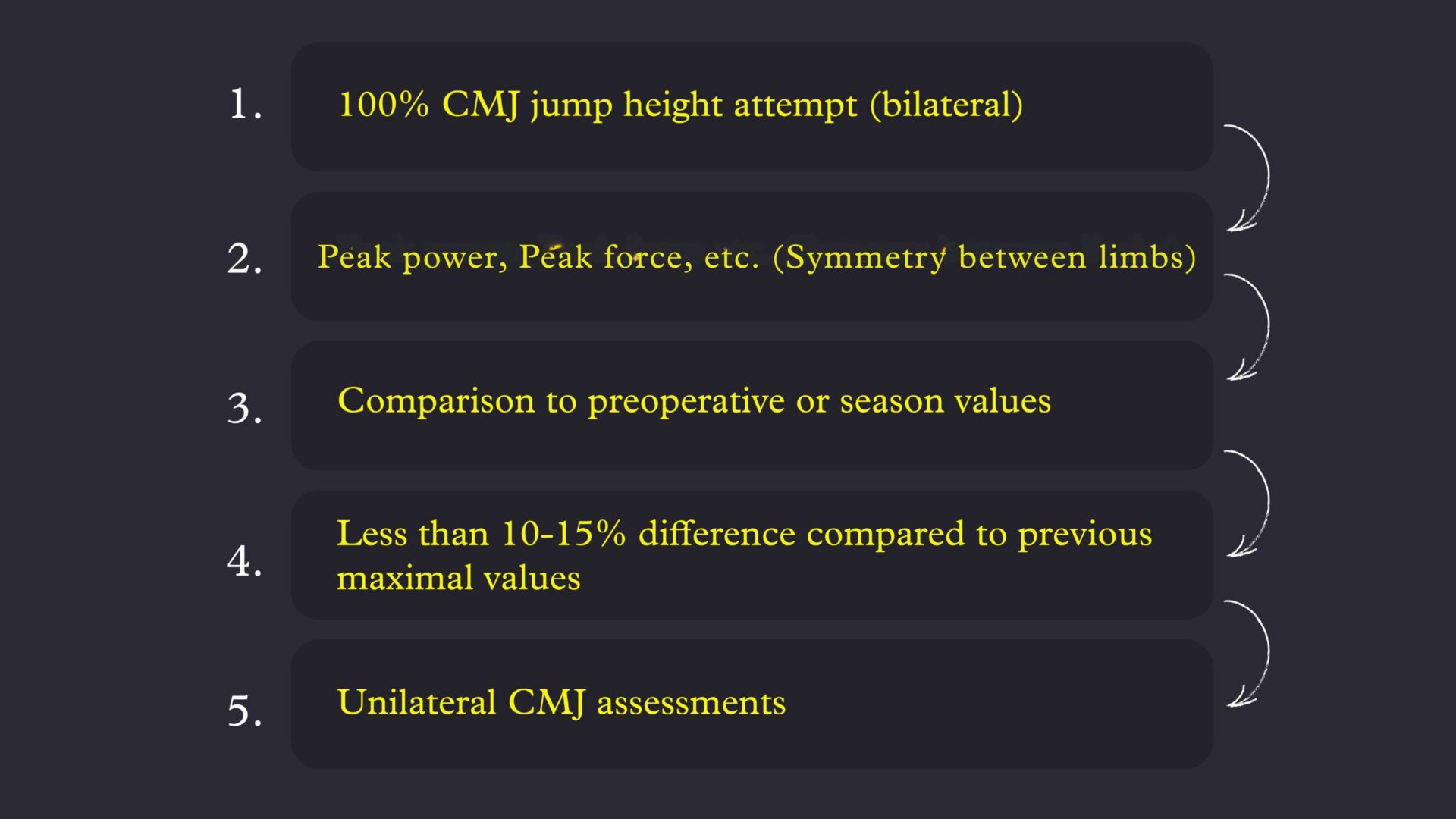
Following successful KAT assessment, the athlete may progress to squat jumps, CMJs, and impact tasks such as drop landings, drop jumps, and single leg exercises.
Notice in the CMJ and drop jump videos that the second landing phase is particularly clean. I interpret it as a suboptimal jump because the athlete is not at 100% of his jump height. If the athlete achieves 100% jump height, the second landing is often not so clean.
Approaching the decision day
These fundamental tests assist in determining whether an athlete may pursue conservative treatment or require surgical intervention.
The athlete’s status is evaluated using four criteria: achieving at least 80% limb symmetry on the 6-meter timed hop, scoring 80% or higher on the Knee Outcome Survey – Activities of Daily Living (KOS-ADL), a Knee Global Rating Score above 60%, and fewer than one giving-way episode in recent weeks. Meeting these benchmarks supports consideration for conservative management.
It is important to note that an athlete’s status may evolve during conservative treatment. Some initially classified as “copers” may later be reclassified as “non-copers.”
These evaluations generate athlete-specific data rather than relying solely on normative datasets found in literature. Each individual possesses unique baseline values, which may not be pathological. For example, a 10% difference between maximal extension torques may be “normal” for some athletes. Emphasis should first be placed on ensuring the validity and reproducibility of measurements, followed by comparisons between limbs, longitudinal tracking of the athlete’s own data, and finally benchmarking against normative values.
Ultimately, the primary goal is to restore the athlete to their personal best performance.
This framework reflects my clinical perspective and requires cautious interpretation. While there are no definitive studies that validate these specific criteria, it offers a practical approach for individualized decision-making.
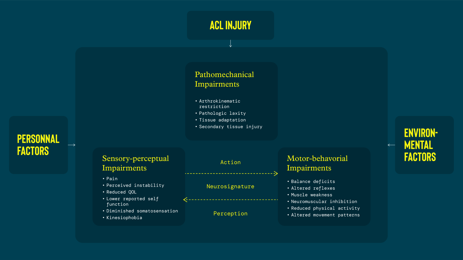
The rationale behind these evaluations is to target the most prevalent deficits commonly observed following an ACL injury. It is essential to acknowledge that additional impairments—particularly in proprioceptive and neurocognitive domains—may also be present.
However, the foundational elements are biomechanical in nature, primarily involving muscle activation, muscle volume, and muscle strength. Without thoroughly assessing and addressing these parameters, the athlete will not have a successful rehabilitation.
Acknowledgements:
Ricardo Andrade for the english reviewing and advice about the writing.
Romain Tourillon, Massamba M’baye and and Simon Barrué-Belou for the clinical and performance inspiration.
Antoine Nordez, Guillaume Le Sant, Lilian Lacourpaille for the supervision during research practice.