Deficits in muscle architecture and tendon mechanical properties are blind spots in rehab. Various tests can quantify reduced force production, poor reactive strength, lower explosive output, and reduced efficiency, but they won’t tell us why the athlete is underperforming. They do not explain that the Achilles tendon is elongating too much, or reveal whether the medial gastrocnemius fascicles have shortened, or if the lateral gastrocnemius is no longer contributing as it should.
Ultrasound steps into these gaps. It helps explain the tissue behaviour driving the performance problem, rather than just the final output.
Ultrasound is not new, but it is newly accessible for sports practitioners.
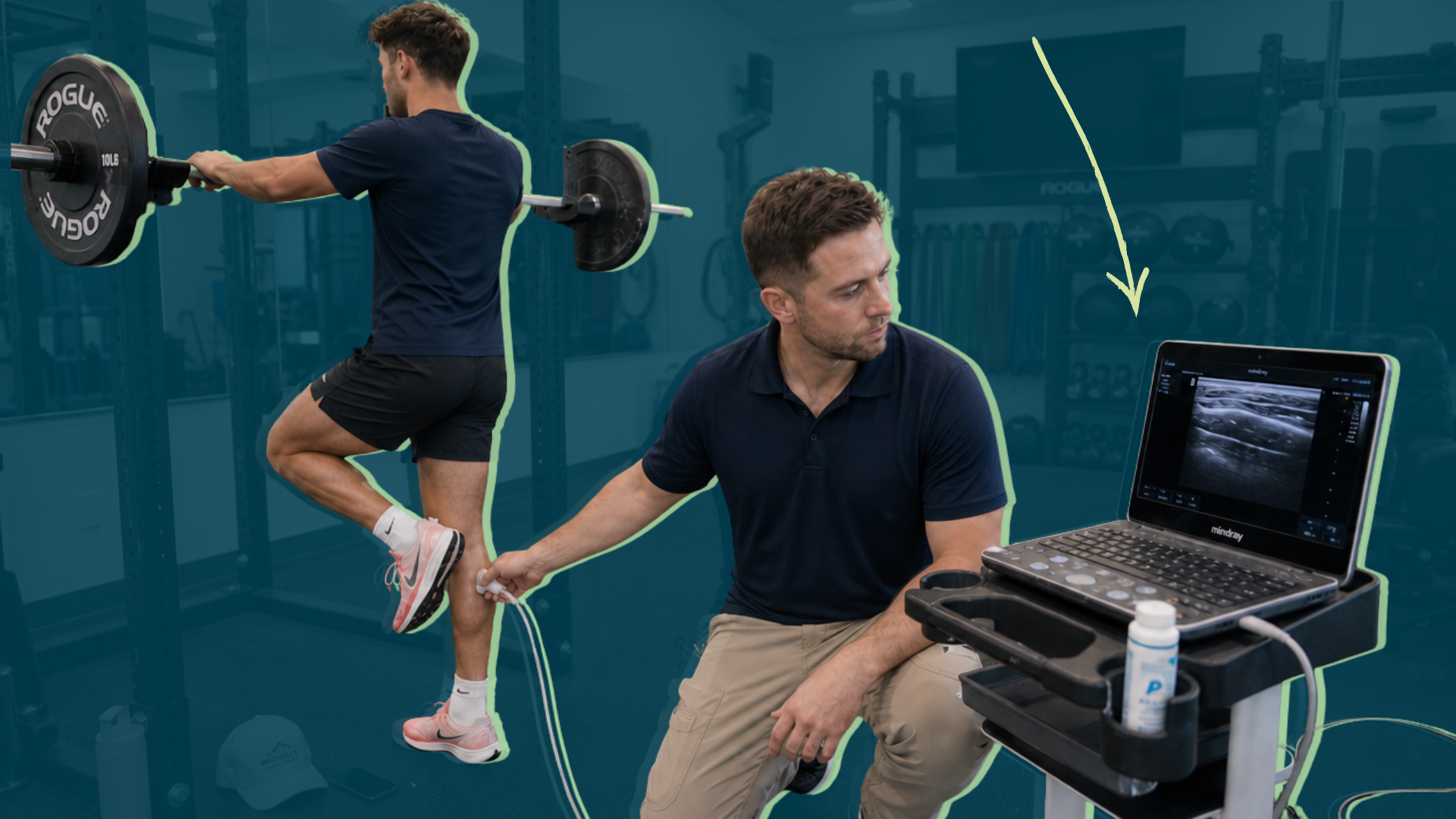
In vivo ultrasound is no longer limited to biomechanics labs and research projects. Portable systems, better image quality, and dynamic probe-fixation options such as Usono’s ProbeFix Dynamic make it possible to use ultrasound on the rehab floor, in the gym, and alongside strength testing. We can now watch muscle fascicles and tendon tissue behave during the tasks we prescribe.
Ultrasound has three particularly useful applications for calf and Achilles rehab. First, ultrasound helps measure muscle architecture and track how it changes during rehab. Second, it helps individualise tendon loading by measuring tendon elongation and estimating strain. Third, it provides useful biofeedback for improving time under tension, targeting a specific muscle or region, or improving movement quality during loading.
Tweet ThisFirst, ultrasound helps measure muscle architecture and track how it changes. Second, it helps individualise tendon loading by measuring tendon elongation and estimating strain. Third, it provides useful biofeedback for improving time under tension.
@colingriffin
Look at what you’re missing
Early ultrasound work in sport science focused mainly on static B-mode imaging. Researchers used it to measure fascicle length, pennation angle, muscle thickness, and tendon behaviour in controlled conditions.[1,2,3,4,5,6] That work mattered because it showed that the triceps surae and Achilles tendon do not behave as one simple unit.
During running, hopping, and jumping, the Achilles tendon can store and release elastic energy while the muscle fascicles operate at relatively small length changes.[3,4] That decoupling is one of the key reasons the triceps surae-Achilles complex is so effective.
Practitioners can now assess more of that behaviour outside the lab. A portable ultrasound, a repeatable setup, and a clear question are often enough to make the tool clinically useful.
With ultrasounds, we can better answer questions like: Is the tendon loading enough to adapt? Is the muscle architecture changing in the intended direction? Is the athlete actually loading the prescribed or desired tissue? Is a good-looking strength score masking a compensatory strategy?
Tweet ThisWith ultrasounds, we can better answer questions like: Is the tendon loading enough to adapt? Is the muscle architecture changing in the intended direction? Is the athlete actually loading the prescribed or desired tissue?
@colingriffin
Measuring muscle architecture
We need more than a peak force number to understand why an athlete has lost plantarflexion function. Muscle architecture shapes force production, contraction speed, and how well the muscle works with the tendon.
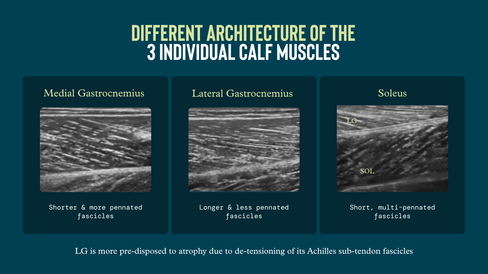
Fascicle length influences shortening velocity and how much contractile range the muscle has available. Pennation angle affects how much contractile tissue can be packed into a given area, and therefore contributes to force capacity. Muscle thickness is a practical proxy for hypertrophy or atrophy.
For the triceps surae, different architectural profiles support different roles. The soleus is built to produce high force repeatedly. The gastrocnemii contribute more to rapid force production and high-velocity tasks. If those architectural features change after injury, performance changes with them. Likewise, because these three muscles have slightly different architectural profiles, we cannot assume the same exercise will load all three muscles in the same way.
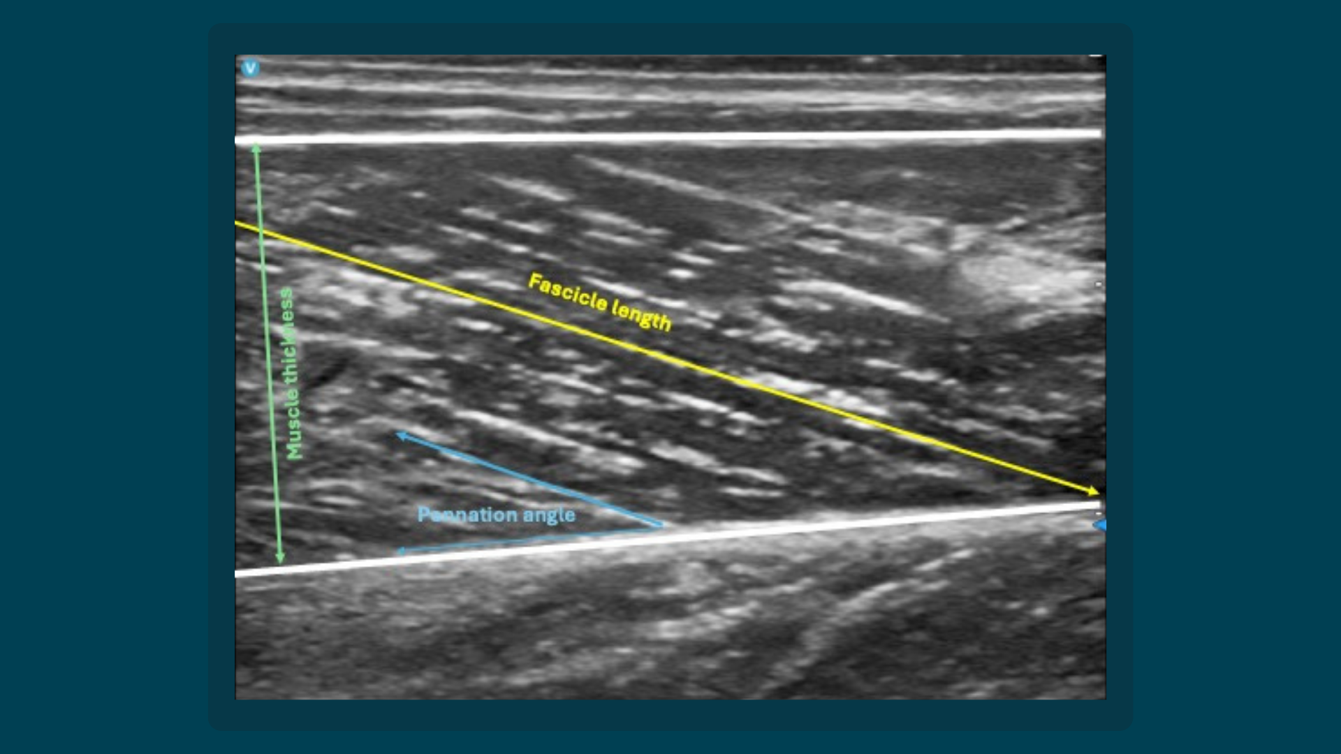
Image the athlete prone with the ankle in a standardised position, typically near neutral, and keep that setup consistent between sessions. For the medial and lateral gastrocnemius, place the probe longitudinally along the muscle belly in order to visualise the superficial and deep aponeuroses and the fascicle path between them. The fascicle itself is not directly visible, but the surrounding perimysial line, which appears white, gives a reliable track for measurement.
The fascicle often runs out of the field of view when imaging muscles with longer fascicle lengths, so we need to extrapolate that value. Consistency matters to ensure repeatable clinical data. A fixed landmark for the soleus is just distal to the lateral gastrocnemius muscle-tendon junction.
Seeing is understanding
While “injury type” matters in rehab, our concern is the impairments that persist.
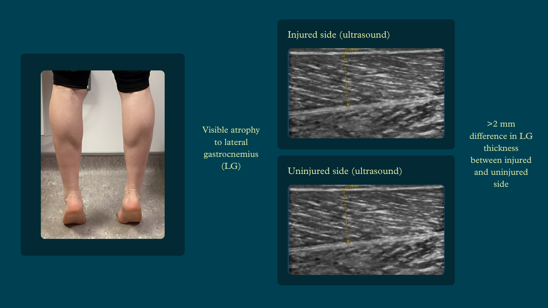
After Achilles tendon rupture, the common pattern is tendon elongation, reduced heel-rise height, atrophy through the triceps surae, and shorter, more pennate fascicles distally. That combination shifts the muscle-tendon unit away from an efficient force-length setup. The athlete can still produce force, although they require greater muscle-tendon unit lengths. But they often lose the end-range plantarflexion capacity and ankle power needed for sprinting, jumping, and reactive tasks.
Achilles tendinopathy often presents differently. Force-sharing shifts across the triceps surae, with the lateral gastrocnemius contributing less in some athletes.[13,14,15] Runners with Achilles tendinopathy show lower neural drive to the gastrocnemius lateralis.[14] Subtendon stiffness is lower in the lateral gastrocnemius than in the other two muscles.[16] This matters clinically, because the athlete may pass a general calf strength screen while still underloading one part of the system.
Medial gastrocnemius strains give another distinct picture. After a calf strain injury there are chronic changes in distal fascicle behaviour, aponeurotic thickening, shorter fascicles, and altered pennation, with reduced EMG in the medial gastrocnemius and increased soleus activation.[8] That often presents clinically as a messy distal medial gastrocnemius image, local scarring between medial gastrocnemius and soleus, and poorer synchronous motion between the two muscle heads in early rehab. Chronic cases may also have fatty infiltration in the distal fascicles and, in some cases, in the mid-belly of the muscle (Video 6).
Tweet ThisAfter Achilles tendon rupture, the common pattern is tendon elongation, reduced heel-rise height, atrophy through the triceps surae, and shorter, more pennate fascicles distally.
@colingriffin
Using exercise to change architecture
Once we know what has changed, we can load with more intent. If we want to target fascicle length, we emphasise long-length isometrics, eccentrics, and partials performed in longer muscle positions.[7,9] For muscle size and force capacity, ensure sufficient load and sufficient time under tension. To bias the distal gastrocnemius, the athlete has to actually work in dorsiflexion and not just move load up and down.
Partial repetitions performed at longer muscle lengths produced greater hypertrophy than partials at shorter muscle lengths, with the advantage particularly evident in central and distal regions, specifically in the gastrocnemius muscles.[9,17] That fits well with what we see clinically in the calf. If the goal is to restore muscle size and architecture in the areas that matter for plantarflexion performance, long-length work deserves a higher place in the programme.
Ultrasound helps verify whether the exercise is doing what we think it is doing.
A football player underwent a plantarisectomy and Achilles tendon debridement for persistent medial Achilles pain.
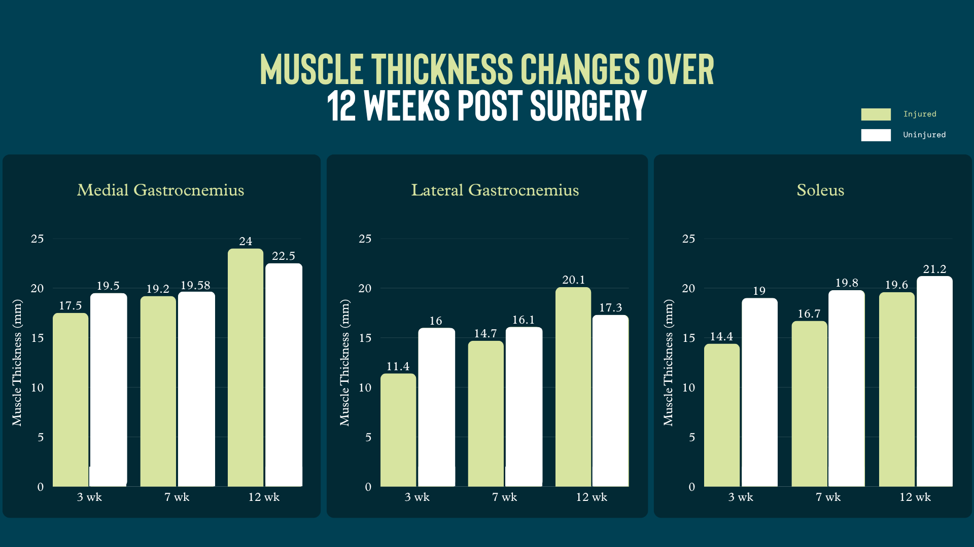
By three weeks post-surgery, ultrasound demonstrated clear muscle size deficits throughout the triceps surae, with the largest reductions in the soleus (~5 mm) and lateral gastrocnemius (~4 mm). The medial gastrocnemius showed a smaller deficit (~2 mm). Rather than simply progressing calf loading according to time, these findings influenced the early rehabilitation priorities. We combined progressive resistance training with blood flow restriction, neuromuscular electrical stimulation, and nutritional support, while using serial ultrasound to monitor muscle thickness throughout rehabilitation.
Over the following 12 weeks, ultrasound showed progressive restoration of muscle size across all three muscles.
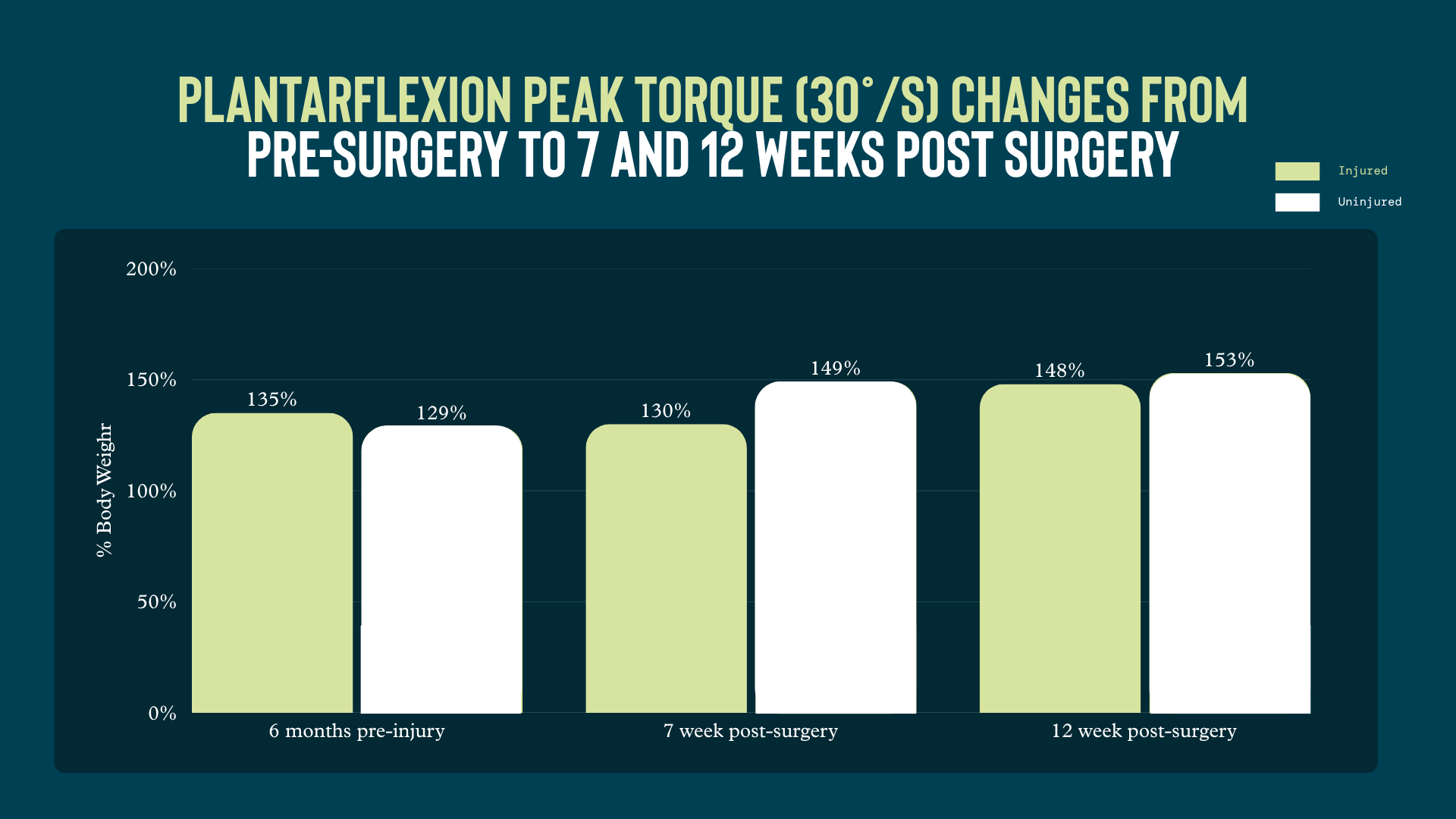
By return to sport, both the medial and lateral gastrocnemius on the injured limb exceeded the contralateral side in muscle thickness, while soleus asymmetry substantially decreased (Figure 4). These structural improvements occurred alongside restored plantarflexor strength, with isokinetic peak torque exceeding pre-injury values by 12 weeks (Figure 5).
Tracking muscle architecture longitudinally gave confidence that we were restoring contractile tissue rather than simply improving performance through compensation, and helped justify progressing load and adding more reactive training.
Measuring Achilles tendon elongation and individualising tendon load
If the Achilles tendon is too compliant, the system loses efficiency. Force transfer slows, elastic return drops, and the fascicles may work harder than they should during running and jumping. If tendon stiffness improves, the athlete usually has a better platform for reactive and explosive output. If a tendon is too stiff, the muscle may lack sufficient strength and its fascicles experience greater eccentric behaviour, which may increase muscle damage and induce DOMS.
It is not enough to know whether the athlete tolerates a calf raise. We want to know whether the tendon was loaded enough to adapt.
The most practical way to determine this is to track displacement of the medial gastrocnemius myotendinous junction during a controlled isometric plantarflexion task.
First, establish resting tendon length. Panoramic ultrasound is one method. Alternatively, mark the top of the free Achilles at the medial gastrocnemius muscle-tendon junction (MTJ) and the distal insertion near the calcaneus, then measure between those marked points in a standardised position.
Keep the probe over the medial gastrocnemius MTJ during loading and record its proximal displacement. Be sure to account for any heel or calcaneal movement: we want tendon elongation and not just total limb movement.
Tendon strain is then:
- Tendon strain = (Tendon elongation / Resting tendon length) x 100
As a simple example, if the resting Achilles tendon length is 20 cm and tendon elongation during isometric loading is 1.1 cm, tendon strain is 5.5%.
That puts the athlete in the adaptive zone. The question is how much external load does the athlete need on the Smith machine or leg press to achieve this strain?
Tendon adaptation appears to respond best to 4.5–6.5% of strain.[10,11,12] Below that, the stimulus may be too small. There may be potential for tendon adaptations up to 9.5%, but this has not been extensively explored. Extremely high tendon strains of >10% may increase tissue stress and accumulate microdamage in an unadapted tendon.
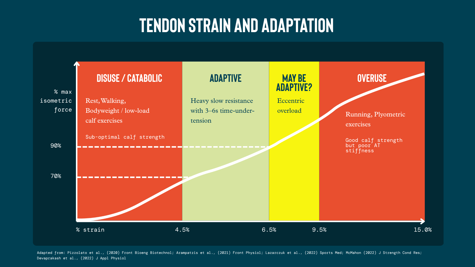
The practical issue is that external load does not result in uniform tendon strain.
Two athletes can both push 80% MVC and achieve different strain values because tendon length, stiffness, muscle force production, and setup all differ.
To transform rehab from a generic loading protocol into a more individual tissue prescription, measure the tendon response, then identify the load that produces useful strain, and build the programme around that.
Zooming out, the Achilles is not one homogenous rope. It is a composite tendon with subtendons linked to soleus, medial gastrocnemius, and lateral gastrocnemius. Force-sharing within the triceps surae changes in Achilles tendinopathy, with the lateral gastrocnemius contributing less in some athletes.[13,14,15]
Tweet ThisTendon adaptation appears to respond best to 4.5–6.5% of strain. Below that, the stimulus may be too small. There may be potential for tendon adaptations up to 9.5%, but this has not been extensively explored.
@colingriffin
The next step is not just “load the tendon more.” It is likely to be “find the correct individual dosage” and “load the right part of the system better.”
With one athlete, we used ultrasound during heavy single-leg Smith machine calf isometrics to identify the external load necessary to achieve approximately 5% Achilles tendon strain. Initially, this occurred at around 1.25x bodyweight during four sets of five 5-second isometric contractions performed in slight dorsiflexion. Rather than progressing load arbitrarily, we reassessed tendon elongation throughout rehabilitation.
Six weeks later, the athlete required approximately 1.5x bodyweight to achieve the same level of tendon strain.
Although we did not measure tendon stiffness directly, the fact that he required substantially greater external load to produce the same tendon elongation provided a clinically useful surrogate for improved tendon stiffness. This gave us greater confidence to progress into higher-velocity calf loading and more intensive plyometric exercises with a tendon demonstrating greater capacity and stiffness qualities.
Time under tension at long muscle lengths
Plenty of athletes can complete calf work without actually loading the tissue in the way we want. They rush the eccentric. They unload the forefoot. They rely more on a passive stretch rather than active tension. Others often rely on “push” isometrics with a floating heel, which does not sufficiently stress the tendon or work the muscle fascicles into length. Dynamic ultrasound helps expose these.
With a live image as biofeedback, the athlete can see whether the fascicle is staying under tension or whether it is shortening early and losing the intended stimulus.
That has been particularly useful for athletes after Achilles rupture, where the soleus often lags despite apparently good compliance with calf work. When they see the difference between a rushed repetition and a controlled one, their performance usually improves immediately.
Targeting regions and individual muscles
Ultrasound also helps bias one region or one muscle. To increase lateral gastrocnemius contribution, direct a straighter knee, work deeper into dorsiflexion, and adjust foot position. A slightly in-toed position can help bias lateral gastrocnemius in some athletes, though it’s still best to confirm it rather than assuming it.
To reduce compensation and restore better medial gastrocnemius contribution, adopt a more externally-rotated foot and use the image to show whether the intended region is actually moving and shortening.
Triceps surae deficits are rarely uniform. One athlete underloads the soleus, while another avoids the lateral gastrocnemius. Another has a local medial gastrocnemius scar that changes fascicle behaviour around the aponeurosis. The image removes the guessing.
Tweet ThisTo reduce compensation and restore better medial gastrocnemius contribution, adopt a more externally-rotated foot and use the ultrasound image to show whether the intended region is actually moving and shortening.
@colingriffin
A professional rugby player has a chronic history of recurrent calf strains involving the medial gastrocnemius and soleus. We detected fatty infiltration in the mid-belly of the medial gastrocnemius. We used dynamic ultrasound to target this region of muscle to stimulate greater activation and remodelling of the neighbouring fascicles in the hope of reducing the fatty infiltration and improving tissue quality.
Building a more complete athlete profile
Ultrasound is most useful when combined with other tools. Strength testing gives the output. Reactive strength testing shows how well that output carries over to fast stretch-shortening tasks. EMG reveals neural drive and muscle contribution, while dynamic ultrasound shows the tissue behaviour underneath those results.
Together, that gives a far better profile than any one test on its own.
An athlete might show reduced reactive strength index, acceptable isometric peak force, and persistent medial gastrocnemius under-recruitment on EMG. Ultrasound then shows poor fascicle behaviour through the distal medial gastrocnemius and low tendon elongation during loading. We can specifically target this and provide the right feedback to the athlete when performing the exercise.
Dynamic ultrasound does not replace performance testing and good clinical reasoning. It adds another layer of precision to assessment and rehab.
Ultrasound helps move beyond output measures to where we can identify the structural and mechanical deficits limiting performance.
Whether monitoring recovery of calf muscle architecture following surgery or individualising tendon loading in an athlete with chronic Achilles tendinopathy, the ability to visualise tissue adaptation allows rehabilitation to become genuinely athlete-specific rather than protocol-driven.
Tweet ThisUltrasound does not replace performance testing. It adds another layer of precision to assessment and rehab. Ultrasound helps move beyond output measures to where we can identify the structural and mechanical deficits limiting performance.
@colingriffin