
The efficiency of pain modulation systems differ with age, physical activity level, and individual characteristics. But the basic neurophysiological mechanisms of pain modulation do not differ between elite athletes and the general population.[1,2]
The biopsychosocial model represents the internationally recognised framework for understanding pain.[3] Nevertheless, in clinical practice—and particularly within the sporting environment—a fundamental component of this model continues to be overlooked and remains inconsistently addressed during the athlete’s initial assessment.[4] Even when clinicians acknowledge the importance of psychosocial factors, they often explore them informally. Instead of a detailed clinical enquiry supported by validated screening tools, they rely on an intuitive determination of the predominant pain mechanisms and key contributing factors.
Clinical judgment alone is not adequate for identifying the elements necessary for an appropriate pain assessment.[5,6] Furthermore, the initial clinical contact can significantly influence prognosis and the transition from acute to persistent pain.[7] An errant perception early in the process can potentially prolong recovery and delay return to sport.
Particularly in elite sport, then, it is difficult to justify neglecting a substantial component of the contemporary definition of pain, thereby failing to characterise the athlete and identifying the principal contributors to their condition.
Tweet ThisAn errant perception early in the process can potentially prolong recovery and delay return to sport.
Alessandro Bazzani
Clinical pain phenotyping distinguishes athletes
Clinical pain phenotyping is the process of identifying and characterising the predominant mechanisms and contributing factors underlying an individual’s pain. It moves beyond biological diagnosis alone to integrate psychological and social domains.[8]
An effective rehabilitation programme must include a multidimensional understanding of the patient’s presentation.
For example, two high level athletes with knee pain and the same clinical diagnosis may require different rehabilitation priorities. One athlete may present with localised pain, and only when the knee is loaded. Their pain is proportionate to the task, they are confident in their recovery, sleep well, show no signs of psychological distress, and have few concerns about returning to sport. In this case, reduced quadriceps strength may be the main factor limiting their progression towards more demanding sporting activities.
The second athlete has the same diagnosis and presents with more widespread pain around the joint. Low load activities easily trigger pain, and the pain feels intense even during relatively simple tasks. The athlete may also be worried about getting injured again, sleep poorly, feel less confident when performing demanding exercises, and expect their recovery to be difficult.
These contrasting presentations raise two important questions: Why can two athletes with the same diagnosis experience pain so differently?, and should their rehabilitation priorities be the same?
Although they share the same pathological diagnosis, factors beyond the anatomical may contribute to these differences and influence what limits each athlete’s recovery.
Psychological and social factors can play an important role. These include both risk and protective factors, such as psychological distress, fear of movement, depression, pain catastrophising, anxiety, recovery expectations, self-efficacy, coping strategies, and the broader social context, all of which are widely recognised as influencing pain and disability.[9,12]
Moreover, growing evidence suggests that the psychological and social factors behind pain presentation may also affect biological processes related to tissue healing,[13] recovery trajectories, and prognosis.[14,15]
Allostatic load is the cumulative burden of psychological and social stressors. A high allostatic load may provide one potential pathway through which these factors influence prolonged recovery, increased pain sensitivity, more persistent swelling, greater discomfort throughout rehabilitation, and greater difficulty progressing training loads. However, despite their potential relevance, only a minority of physiotherapists—and an even lower proportion of physicians— routinely assess psychosocial factors.[4,10,11]
How psychosocial factors influence pain and recovery
The cumulative physiological burden associated with persistent psychological and social stressors can influence pain and recovery through neural, neuroendocrine, cellular, and behavioural mechanisms. These stressors can increase pain sensitivity through alterations in descending inhibitory pathways and nociceptive modulation mechanisms.[17,18] This means that the nervous system may become more responsive to sensory input, potentially resulting in pain being experienced more easily, widespread, or more intensely.
Persistent psychological and social stressors can disrupt the hypothalamic–pituitary–adrenal axis, one of the body’s main systems for coordinating the physiological response to stress. This can alter the regulation of cortisol, a hormone involved in the stress response; and increase the production of reactive oxygen species. These are molecules that, when produced in excess, can cause oxidative stress and damage cells. This physiological and cellular stress may also promote stress-induced premature cellular senescence[16], a process in which prolonged stress causes cells to enter an aging-like state earlier than they normally would.
These neuroendocrine and cellular processes can contribute to a pro-inflammatory biological environment. They may influence pain processing, increasing sensitivity by lowering the activation threshold of peripheral nociceptors, making them more responsive to sensory input. Inflammatory signals can also affect pain modulation within the central nervous system by altering the balance between descending inhibitory and facilitatory mechanisms that regulate nociceptive input.[19]
Beyond these potential neuroendocrine and inflammatory mechanisms, behavioural factors may also affect recovery. Movement avoidance, reduced confidence in loading the affected area, and adopting protective motor strategies may contribute to slower functional progression and make it more difficult to progressively restore physical capacity and key performance benchmarks.
Clinical pain phenotyping enables clinicians to integrate information from the biological, psychological, and social domains in order to understand the mechanisms underpinning pain and disability. Through this process, clinicians can identify the predominant pain phenotype and the potential involvement of pain sensitisation.
This information can guide clinical reasoning, treatment planning, and the development of a shared explanatory model of pain with the athlete: “Making Sense of Pain.”
Why identifying the pain phenotype matters
Many healthcare professionals tend to view pain primarily in its acute form, where tissue injury or overload is associated with the onset of symptoms. In such cases, pain typically decreases over time as biological healing occurs, ultimately leading to symptom resolution and restoration of function.
However, this represents only one of several possible clinical presentations. Pain is “an unpleasant sensory and emotional experience associated with, or resembling that associated with, actual or potential tissue damage.”[3]
Therefore, when assessing an athlete with pain, it is essential to consider the predominant pain type (i.e., the underlying pain mechanisms), its characteristics (mechanical or non-mechanical symptom behaviour), and the degree of sensitisation (i.e., the level of nervous system responsiveness and modulation efficiency).
Pain sensitivity is a clinical umbrella term for how some individuals experience and perceive pain to a greater extent than others. It can be explored through clinical sensory testing, using standardised stimuli such as light touch, pinprick, pressure, cold, and temporal summation. Importantly, these findings indicate the presence and extent of pain sensitivity, but do not identify the specific neurophysiological mechanisms responsible for it.[20]
A multidimensional assessment helps explain why, in some cases, pain may appear disproportionate to the extent of tissue injury, why symptoms may persist beyond expected healing timelines, or why swelling may persist for a prolonged period.[21,22]
Furthermore, identifying the predominant pain phenotype can guide treatment selection: rehabilitation, pharmacological, behavioural, or surgical.[23] Recent phenotype-informed care models further suggest that the intensity, duration, and multidisciplinary composition of care may be titrated according to the complexity of the clinical phenotype, rather than being determined by the biological diagnosis alone.[24]
It is also important to recognise that certain medications are specifically indicated for particular pain conditions, such as neuropathic pain, while other interventions specifically target pain sensitisation and the increased excitability of the nervous system that contributes to the pain experience.[25]
Integrating pain phenotyping into clinical practice
There are several frameworks that clinicians can use to facilitate pain phenotyping. Less important than adopting a specific framework is assessing pain as a multidimensional experience.
Validated screening tools are crucial components of the assessment process. Without systematically collecting this information, clinicians risk relying solely on clinical impression, thereby reducing the accuracy of athlete phenotyping.
The Örebro Musculoskeletal Pain Screening Questionnaire (Short Form) is a practical screening tool that easily integrates into routine clinical practice. It consists of 11 items, each scored on an 11-point scale (0–10), assessing multiple domains associated with the risk of persistent pain and disability, including pain intensity, perceived function, psychological distress, depressive symptoms, recovery expectations, and fear avoidance beliefs.[27]

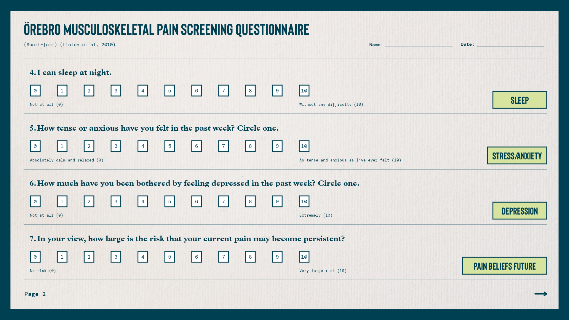

Analysing individual responses also allows clinicians to identify specific domains requiring further investigation. For example, if a clinician identifies elevated levels of psychological distress, the Depression, Anxiety and Stress Scale (DASS-21)[28] may help determine which emotional factors are predominantly influencing the athlete’s presentation. Similarly, when the athlete exhibits high levels of pain catastrophising or fear avoidance behaviours, instruments such as the Pain Catastrophizing Scale (PCS)[29] or the Pain Self-Efficacy Questionnaire (PSEQ)[30] may further support clinical reasoning by helping target rehabilitation strategies and providing useful outcome measures for monitoring progress throughout rehabilitation.
This approach facilitates a more individualised rehabilitation programme, and helps determine whether referral to other healthcare professionals, such as a psychologist, may be appropriate within a multidisciplinary management pathway.
Within elite sport, systematically overlooking these domains means disregarding clinically relevant information that may influence prognosis, treatment adherence, and return to sport outcomes. If pain is multidimensional, its assessment must also be multidimensional. Without a structured process of clinical pain phenotyping, clinicians risk treating only the biological diagnosis rather than the athlete experiencing pain.
Tweet ThisIf pain is multidimensional, its assessment must also be multidimensional.
Alessandro Bazzani
Semi-pro soccer player case study
A 26-year-old semi-professional male soccer player, playing as a central midfielder, presented with a six-month history of persistent left sided groin pain. He trained four times per week and competed in one match per week, while also working three shifts per week as a bartender. His primary concern was his inability to return to play, accompanied by increasing anxiety regarding contract renewal and the possibility of not securing a contract with another team.
Symptoms began approximately six months before presentation during the preseason, following an increase in training load and a transfer to a new club. The athlete perceived this as a period of considerable performance-related pressure.
Initially, pain was mild—3/10 on the Visual Analogue Scale—localised to the left groin, and primarily provoked during changes of direction to the right. Over the following weeks, symptoms progressively worsened: he scored them as 8/10 on the VAS, the pain became more diffuse, and it ultimately prevented his participation in training and competition.
During the months preceding the assessment, pain also began to interfere with activities of daily living, including walking, stair climbing, and prolonged sitting. Concurrently, increasing stress and anxiety related to uncertainty about his sporting career developed, while symptoms gradually spread along the medial thigh towards the knee.
The athlete initially underwent conservative management consisting of massage therapy, dry needling, and an adductor strengthening programme. Despite treatment, he reported no clinically meaningful improvement and experienced repeated symptom exacerbation whenever attempting to return to training.
Imaging revealed CAM morphology, femoroacetabular impingement, and an acetabular labral tear. Based on these findings, the orthopaedic surgeon proposed hip arthroscopy.
Psychosocial assessment
The athlete scored 71/100 on the Örebro Musculoskeletal Pain Screening Questionnaire (Short Form), indicating a high risk of future disability (>50). The domains most affected were recovery expectations, stress, and anxiety. These findings were further supported by the Depression, Anxiety and Stress Scale (DASS-21), which demonstrated severe levels of stress and anxiety.
From a cognitive perspective, the athlete demonstrated strong biomedical beliefs. He attributed his pain primarily to the MRI findings (CAM morphology, femoroacetabular impingement, and labral tear), a belief reinforced during previous medical consultations.
From a social perspective, soccer was his primary source of income, resulting in considerable pressure to return to competition as quickly as possible. He also reported poor sleep quality.
Physical examination
Clinical examination revealed marked tenderness on palpation of the adductor conjoint tendon and pubic symphysis. He experienced pain during both the adductor squeeze test and the crossover test, with both activities also reproducing the athlete’s familiar radiating symptoms into the medial thigh. In contrast, femoroacetabular impingement provocation tests and labral tests did not reproduce the athlete’s familiar symptoms.
Clinical pain sensitivity was also part of the physical examination. The athlete reported pain intensity of 8/10 on the VAS in response to an ice cube enclosed in plastic on the symptomatic region for 10 seconds. This indicated marked cold pain sensitivity. The widespread symptom distribution, symptom persistence, and exaggerated responses to provocative testing all contributed to an increased sensitisation profile.
Functional assessment
Movement analysis during change of direction tasks demonstrated altered movement strategies: increased anterior pelvic tilt, greater hip flexion, increased lateral trunk lean, and greater trunk rotation. These were all associated with increased symptom provocation.
Strength and performance assessment demonstrated reduced hip abductor strength on the affected side, impaired trunk control during anti-rotation and anti-extension tasks, reduced plantarflexor strength, and a decreased Reactive Strength Index (RSI).
Overall, the functional assessment identified specific physical capacity and movement control deficits that appeared to reduce tolerance to sport-specific change of direction loads.
Clinical reasoning
The diagnosis was adductor-related groin pain presenting with features of central sensitisation and secondary hyperalgesia.
We did not consider the imaging findings of CAM morphology, femoroacetabular impingement, and a labral tear clinically relevant because they were inconsistent with the clinical presentation, and commonly occur in asymptomatic populations.[31,32]
The athlete’s pain was a mix of both nociceptive and nociplastic features, with evidence of increased pain sensitivity. The consistent reproduction of familiar symptoms during local palpation and adductor loading supported the nociceptive contributiong. However, symptom persistence and progressive spreading, repeated exacerbations, and an increased pain response to cold stimulation suggested altered nociceptive processing and a sensitised pain presentation.
| Domain | Risk / contributing factors | Protective factors |
| Biological / physical | Hip abductor weakness, reduced plantarflexor strength, impaired trunk control, reduced RSI, altered COD strategies | Young age; good aerobic conditioning; preserved quadriceps and hamstrings strength |
| Psychological (affective and cognitive) | Elevated stress and anxiety, dysfunctional biomedical beliefs, poor recovery expectations | Motivation to return to sport; openness to alternative explanations of pain |
| Social | Contract renewal pressure; financial dependence on sport | Access to multidisciplinary care |
| Lifestyle | Poor sleep quality | Appropriate nutrition behaviours; absence of other major lifestyle stressors or comorbidities |
Taken together, these findings suggested that an intervention based exclusively on the biomedical diagnosis and the identified physical impairments would have failed to adequately account for the intensity and widespread nature of the athlete’s symptoms. Integrating psychosocial screening shaped a more comprehensive pain phenotype, and facilitated a more accurate shared understanding of the athlete’s pain.
Multidisciplinary approach to management
Management consisted of pain education, progressive exercise rehabilitation targeting the deficits identified during the assessment, and an individualised load management programme.
Given the prominent psychosocial contributors, we emphasised multidisciplinary management. A sports psychologist came on board to address stress, anxiety, and poor sleep quality, while the coaching staff collaborated to provide a supportive environment throughout rehabilitation.
Determining an athlete’s clinical pain phenotype and related characteristics a priori, without first conducting a systematic assessment of psychosocial factors, represents a significant clinical bias that may directly lead to misguided clinical decision-making. In such cases, clinical reasoning is replaced by assumptions, often driven by subjective impressions or a reductionist interpretation of the problem. Consequently, rehabilitation planning and pain management decisions are built on weak, if not arbitrary, foundations.
Without the process of pain phenotyping, any conclusion regarding an athlete’s pain is incomplete and potentially misleading for both clinical management and return to sport decision making. This increases the risk of misunderstanding the clinical presentation and adopting inappropriate management strategies, ultimately contradicting the principles of contemporary pain medicine.
Tweet ThisIn such cases, clinical reasoning is replaced by assumptions, often driven by subjective impressions or a reductionist interpretation of the problem.
Alessandro Bazzani