Working in professional sport, we are familiar with the scenario of rehabilitating an athlete with an injury that has put their career in jeopardy. I have referred to an ACL injury for a footballer as a “catastrophic injury.” My outlook gained a new perspective when I led the rehabilitation of a professional goalkeeper who suffered a life threatening cervical spine (CSp) injury during a road traffic accident (RTA).
Her initial aim was to achieve basic quality of life. She went on to return to playing professional football.
Most of my career has been as a physiotherapist in professional football. I now work privately at Rehab 4 Performance (R4P) Sports Injury Clinic in Liverpool, UK, where I specialise in managing lower limb injuries. Upper limb injuries are infrequent, and spinal trauma practically unheard of. Achieving the hoped for outcome for this athlete imposed a steep learning curve on me.
This case study catalogues her clinical journey, and my own journey as I drew on my professional network to fill knowledge gaps both in terms of assessment and rehabilitation.
Tweet ThisMy outlook gained a new perspective when I led the rehabilitation of a professional goalkeeper who suffered a life threatening cervical spine (CSp) injury during a road traffic accident (RTA).
@matt_kono
Pathology and acute management
The mechanism of injury was ejection from the back seat of a car through the front windscreen, on to the side of the road. She suffered numerous significant injuries that affected both the axial and appendicular skeleton. This case study will focus on the head and neck injuries, to include the right shoulder.
Jefferson “burst” fracture
The Jefferson fracture refers to a burst fracture of the atlas (C1) resulting in double fractures of the anterior and posterior arches. The mechanism of injury commonly involves axial compression forcing the occipital condyles into the lateral masses of the atlas.
Given the damage to the transverse atlanto-axial ligament, the injury was considered unstable and a halo device was implemented to avert spinal cord injury. A halo device consists of a ring that is attached to the skull with pins. A vest is attached to the ring with rods.
Concomitant injury to the axis (C2) is typical, and this player incurred three further fractures here.
Post-concussion syndrome
Over 30% of all concussions will go on to having long-standing symptoms. [1] Headache, dizziness, fatigue, sleep disturbance, and concentration and memory problems are all examples of ongoing symptoms.
The Amsterdam Consensus Statement deems symptoms lasting beyond four weeks as “persistent.” [2] A multi-modal clinical assessment is recommended.
The pathophysiology is unknown, but the main theories are: autonomic nervous system instability (i.e., blood flow), metabolic / inflammatory, visual / vestibular dysfunction, and cervical spine dysfunction.
We do not know which of these theories is most linked to post-concussion syndrome, let alone the athletic population specifically. This athlete had suffered one previous concussive episode. However, the potential long term sequelae from repetitive impacts is not clear due to methodological variability. [2]
Tweet ThisThe mechanism of injury was ejection from the back seat of a car through the front windscreen, on to the side of the road. She suffered numerous significant injuries that affected both the axial and appendicular skeleton.
@matt_komo
Initial presentation for our rehab
The athlete was 24 years old and attended rehabilitation for eight months after the accident with an Aspen collar before the work described here. That earlier rehabilitation had been limited to compound bodyweight exercises and steady state bike sessions that we had prescribed remotely.
She had a relatively unremarkable injury history prior to the accident, aside from the concussion mentioned above.
Cervical spine and shoulder
An x-ray immediately following the road traffic accident demonstrated the absence of any injury to the bones of the shoulder. However, given the enormity of her CSp injuries, the shoulder symptoms had not yet been addressed.
Overhead activity and lying on her right side provoked shoulder symptoms. She experienced instability in the shoulder, which she described as “looseness.”
Objectively, there was atrophy of the posterior rotator cuff muscles and a flexion deficit of 40-50° due to antero-superior gleno-humeral joint pain. Posterior cuff force production was weak. Apprehension testing was positive. Palpating the long head of the biceps caused some soreness. Rotator cuff improvement tests restored full pain-free shoulder flexion, implying that enhanced recruitment of these muscles was the stability platform to facilitate range of movement in the absence of symptoms.
Considering her neck injuries and subsequent loss of function due to immobilisation, we could not rule out pain referral from the CSp. However, given the clinical presentation, our working hypothesis for the shoulder was anterior instability secondary to trauma, and we managed this conservatively.
The athlete had profound CSp muscle atrophy, and the associated weakness and loss of range of motion (Table 1). Simply holding her head upright was a challenge. She described a constant background ache in the right sub-occipital region, with an intermittent sharp pain on sudden movements.
| Movement | Athlete ROM(°) | Comments | Benchmark ROM(°) |
| Flexion | <5 | Negligible head-on-neck motion Primarily lower CSp motion | 80 |
| Extension | 20 | Negligible head-on-neck motion Primarily lower CSp motion | |
| Lateral flexion | 5 | Lower CSp motion only Limited by contralateral tightness in the scalenes and upper fibres of trapezius musculature. | |
| Rotation right | 20 | Not clean rotation; combination of flexion and lateral flexion Overactive sternocleidomastoid | 80 |
| Rotation left |
Tweet ThisOverhead activity and lying on her right side provoked shoulder symptoms. Objectively, there was atrophy of the posterior rotator cuff muscles and a flexion deficit of 40-50° due to antero-superior gleno-humeral joint pain.
@matt_komo
Post-concussion syndrome
At her initial examination, the athlete had the following symptoms associated with PCS: headaches, vertigo, short term memory loss, photosensitivity, and diminished concentration.
Neurological examination did not raise any concerns regarding post-concussion syndrome (PCS). Normally, we would have performed a graded aerobic test such as the Buffalo Treadmill Test to determine whether symptoms were associated with autonomic instability or physical deconditioning. [3, 4] This was not appropriate at this stage given the spinal injuries. However, we began challenging the aerobic system incrementally using the Watt Bike.
Her Vestibular-Oculomotor Screening Assessment was normal, so we approached the PCS as being cervicogenic.
The cervical spine protects neural pathways associated with coordination of head and eye movement, posture, and balance. Cervicogenic headaches are well evidenced. There is a correlation in symptoms and management between whiplash associated disorder and concussion: exercise and manual therapy provide favourable outcomes for both. [5]
Education and reassurance are important. [3] We explained to the athlete that the persistent symptoms did not necessarily reflect ongoing physiological brain injury but instead were linked to factors relating to her CSp injuries. The athlete was reassured that addressing these factors would likely address her PCS symptoms. Psychological support was provided by the athlete’s employer.
Known knowns, and known unknowns
In my current and previous leadership roles, it is vitally important to acknowledge the strengths and weaknesses of the multi-disciplinary team, including my own. This let us direct the athlete to the relevant practitioner to facilitate optimal outcomes for both the athlete and the organisation. Of equal importance is identifying the learning requirements of the staff, formulating and then implementing a strategy to address them.
Before this, I had not led case management, nor had I been involved in the rehabilitation of an individual with CSp trauma of this magnitude. Once I acknowledged this gap in my learning and experience I accessed support from the MDT at R4P and consulted my wider professional network. This included the neurosurgical team managing the athlete’s CSp injuries, a sport and exercise medicine consultant, a shoulder physiotherapy specialist, senior rehabilitation physiotherapists in professional Rugby Union, and academic researchers at Coventry University.
Our sport and exercise medicine consultant had extensive experience with concussion management, and provided medical oversight for the rehabilitation. I also embarked on a comprehensive concussion course — Complete Concussion Management — that included concussion pathophysiology, the autonomic nervous system, cognitive function, baseline and post-injury concussion testing, acute injury assessment, and how to address complex persistent concussion symptoms. We worked closely with the athlete’s neurosurgeon to ensure sufficient fracture healing and a stable atlanto-axial prior to commencing or advancing CSp loading.
I was extremely familiar with was the rehabilitation process for athletic injury. Best practice requires a criteria-based approach using benchmarked objective data to guide progression and return to function.
First, we must determine which physical capabilities someone needs to achieve and why; the tests which assess those qualities and how they are performed; and the meaning of benchmark(s) for each test.
Tweet ThisIt is vitally important to acknowledge the strengths and weaknesses of the multi-disciplinary team, including my own. This let us direct the athlete to the relevant practitioner to facilitate optimal outcome.
@matt_komo
Strength and stiffness during CNI
The level of contact and the diving actions associated with goalkeeping expose the CSp to high impact forces. The local musculature must be capable of absorbing these forces to promote physical performance and mitigate injury.
Contrary to popular opinion, strength does not appear to mitigate concussion unless the individual is aware of the impending impact. [6, 7] The time it takes to activate the neck muscles volitionally is greater than the amount of time necessary to induce a concussion secondary to impact. [8]
Fatigue may be a contributing factor to concussive injury after multiple impacts. [9] Stronger muscles may be more resistant to the negative effects of fatigue induced by repeated actions.
Greater neck stiffness, achieved by preloading muscles, facilitates greater energy absorption after impact. Research with collegiate American football players concluded that muscle stiffness, not strength, reduces the number of severe head impacts in a season. [10] This may mitigate concussion and should be part of rehabilitation following CNI.
Assessing cervical spine muscle strength
Given the profound weakness, initial assessment and rehab used pressure biofeedback. However, this was not sufficient to assess force production of the CSp musculature in a way that could inform a return to elite sport.
Tools like an isokinetic dynamometer or force plates were not appropriate, but colleagues from a Premiership Rugby Union team — a sport where CSp trauma is more common, and concussion is the most frequent match injury [11] — directed me to the Gatherer System (GS). The GS permits objective strength testing for the neck in validated positions, and functions as a dynamic training tool.
Early stage strength assessment with pressure biofeedback starts with inserting the folded cell beneath the neck just below the occiput. For deep neck flexor assessment, instruct the athlete to nod their head with minimal activity in the superficial neck flexors.
During later stages of rehab, we used the GS for an isometric break test procedure. The exact setup varied according to the muscle group. However, we maintained the reliability of the setup.
Benchmark data for the Gathering System
It is not unusual when rehabilitating elite athletes to encounter situations where there is no perfect data set or robust evidence for a particular test or intervention. Performance and medical teams are often creating the evidence, given the unique population and environment they interact with.
There was no data set available for CSp muscle strength in elite goalkeepers, let alone females. As such, we referenced benchmark data from a Premiership Women’s Rugby Union team (Table 2). Acknowledging anthropometric differences between positions in rugby union, we used data representative of the “backs.”
| Movement | Force (kg) |
| Flexion | 17-22 |
| Extension | 30-42 |
| Lateral flexion | 28-38 |
CNI rehabilitation
Given the cervicogenic nature of the PCS, we approached the post-concussion rehabilitation alongside that of the CNI, rather than each in isolation. Figure 1 shows the interconnected relationship and basic hierarchy between physical components.

Range of motion
We started working on range of motion (ROM) on my first day with the athlete, initially with support to eliminate the effect of gravity.
Local soft tissue techniques helped reduce muscle shortening that had resulted from halo immobilisation, and also to target symptoms associated with the PCS, particularly cervicogenic headaches. Structures innervated by the C1-C3 nerve roots—including the sub-occipital muscles, splenius capitis, and upper trapezius—are commonly associated with cervicogenic headaches. [12]
Low load resistance training
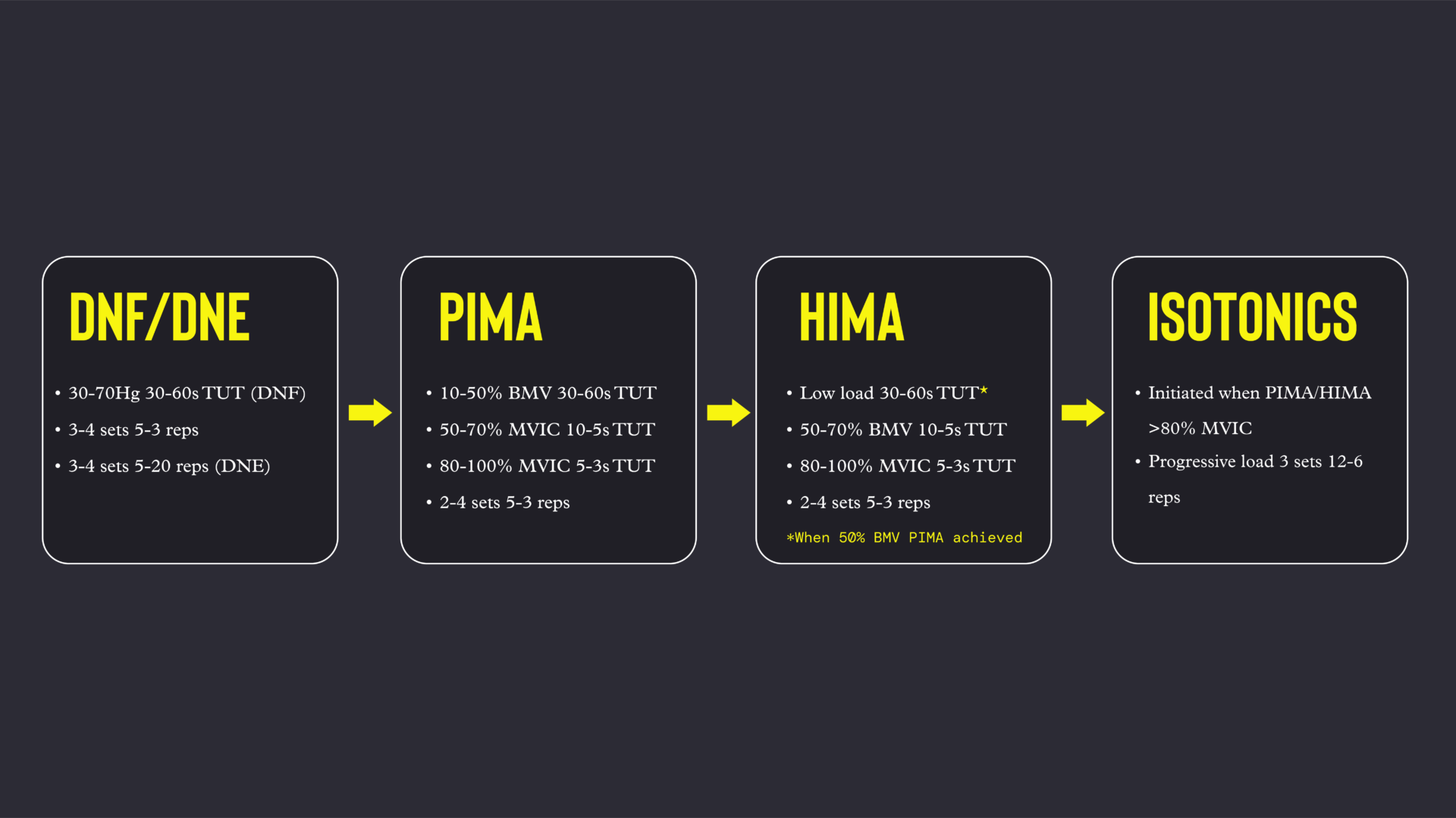
Figure 2 demonstrates the loading continuum for the CSp musculature. Low load resistance training also began on Day 1.
Training of the anterior neck musculature began with cranio-cervico (head on neck) flexion initiated by the deep neck flexors (DNF). Individuals with neck pain find it difficult to perform this action, particularly if they are weak following immobilisation. Given her loss of range of motion, the ROM training facilitated the early resistance work.
Practitioners can monitor overactivity of the superficial sternocleidomastoid muscles by palpating the structures.
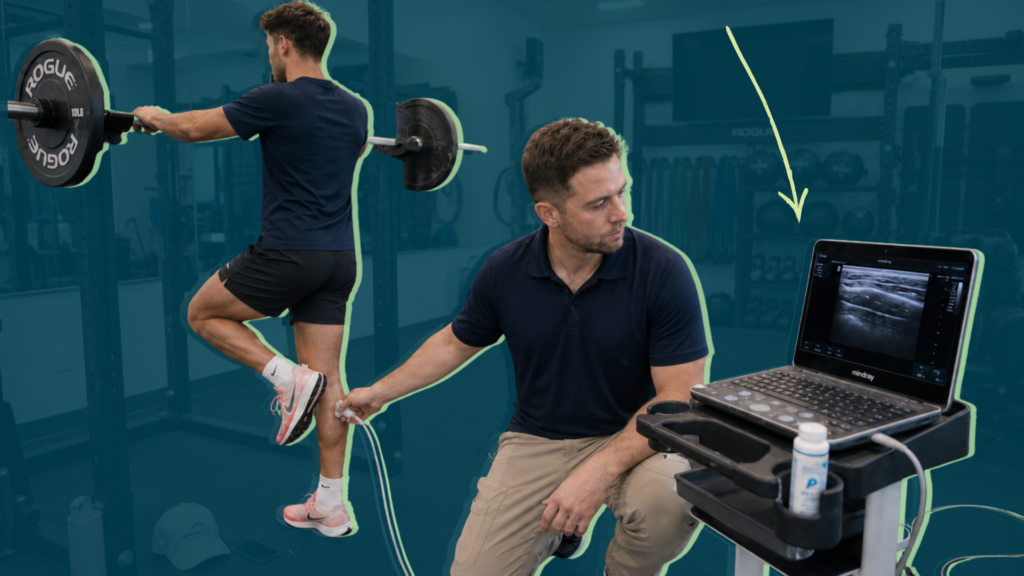
Pressure biofeedback, progressing from 30 to 70 mmHG and 5 to 60 seconds time under tension, rebuilt the capacity of the DNF muscles. Nodding and holding at 20° and 40° CSp flexion from the horizontal helped ensure activity of both the DNF and superficial neck muscles (SNF), such as the sternocleidomastoid and scalenes.
Training for the CSp deep neck extensors (DNE) commenced in the four-point kneeling position. Cranio-cervico extension was facilitated before independent performance. Repetitions progressed from five to 20. A laser pen attached to the head facilitated rotation; and external cueing such as imagining a “skewer through the middle of the head” promoted clean rotation without unwanted movement.
Palpating the lower CSp and ensuring proper motion facilitated lower cervical extension with retraction.
Tweet ThisTraining of the anterior neck musculature began with cranio-cervico (head on neck) flexion initiated by the deep neck flexors (DNF). Individuals with neck pain find it difficult to perform this action, particularly if they are weak following immobilisation.
@matt_komo
Proprioceptive training
Proprioception, in particular joint position sense (JPS), is pivotal alongside strength training for symptoms associated with cervicogenic PCS and CNI. The CSp is rich in golgi tendon organs, muscle spindles, and joint receptors. JPS training started in week 2, when she had developed sufficient ROM. We mounted a laser on the player’s head as she sat facing a target board one metre away. She rotated her head to either side before returning to the centre, initially with eyes open. We applied the same idea to extension.
The exercise progressed by her closing her eyes before moving the head away and back towards the centre. Another possible progression would be increasing velocity of movement. Tracing or following various free-form patterns with the laser challenged fine motor control with combined movements.
Isometric resistance training
She achieved the DNF and DNE objectives in week 4, opening the door to isometric training with the GS. Isometrics started with pushing isometric muscle actions (PIMAs) in a neutral cervical spine position to mitigate axial loading and shear forces. The PIMA permits the athlete to control the intensity of the force production, whilst the GS trainer provides objective force data via a load cell to inform progression / regression. The setup followed the testing protocol.
The peak isometric force for the first session was equal to or less than 10% of the lowest benchmark value associated with the movement (Table 2), e.g., flexion started at 1.7kg. We progressed intensity at low levels by increasing time under tensions from 30 to 60s.
When the player reached 50% of the BMV, her maximum voluntary isometric contraction on the GS trainer became the basis for intensity (Figure 3). This permitted safe load progression relative to the athlete’s force production capability.
Holding isometric muscle actions (HIMAs) began when she produced 50% of BMV with a PIMA. This was about three months into our rehabilitation together. This allowed the athlete to build confidence as well as load tolerance before performing an eccentric contraction where she would not self-select the force, but instead would respond to an external load.
Loading started with very light loads for durations of 30-60s, progressing relative to BMVs until 70%, at which point maximum voluntary isometric contraction break testing guided load progression. This relatively aggressive test loads the CSp beyond its force absorption capabilities, hence the decision to use BMVs as a guide up to 70% BMV.
Tweet ThisHolding isometric muscle actions (HIMAs) began when she produced 50% of BMV with a PIMA. This was about three months into our rehabilitation together. This allowed the athlete to build confidence as well as load tolerance.
@matt_komo
Isotonic resistance training
When MVIC actions reached 80% (about five months in), we started isotonic muscle contractions at low intensities, with the neurosurgeon’s approval (Figure 2). This ensured that she had a good base level of strength prior to loading the CSp through range with increasing compressive and shear forces. [13] The beginning of resistance training was also when we began combined movements.
Stiffness training
Reactive stiffness started alongside GS break testing at 70% BMV, six months after beginning our rehab journey.
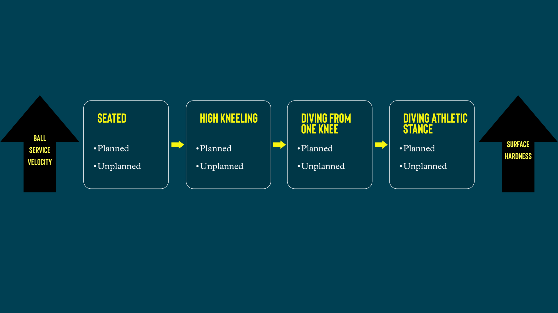
We initially trained stiffness in non-functional positions, then went on to train unplanned stiffness in more football specific positions related to the positional requirements of a goalkeeper. We progressed the intensity of loading using resistance cords of varying strength and velocity of external loading.
Diving continuum
This article is only recounting our work with this player’s cervical spine injuries — we’re not going into the concurrent injuries and her conditioning. However, it is important to highlight that lower limb and central conditioning ran alongside the CNI specifics described in this article, each with their own respective benchmarks. These physical capabilities were important to establish readiness to cope with the demands of diving, linking a strength and stiffness foundation in the lower limbs and torso with that of the neck.
Once she reached 80% BMV with the GS break test (6.5 months), we commenced the diving continuum (Figure 3).
To mitigate impact forces and foster confidence, we used techniques like diving from a high kneeling position and landing on cushioned surfaces. We programmed planned and unplanned variations to challenge muscle stiffness to varying degrees. She handled incoming balls at progressively increasing velocities.
This was a landmark moment in the rehabilitation process for the athlete. Doubts about whether she would ever be able to be a goalkeeper again evaporated.
Understand your weaknesses to build a stronger team
High impact, combative sports such as rugby and American football have led the way in terms of profiling CSp strength, rehabilitation, and injury mitigation. As a wider consideration, there is a strong argument for football medical and sports science departments to become more proactive with CSp strength endurance and reactive stiffness as we strive to mitigate CSp injury and concussion.
As an external provider and given the severity of this rehabilitation, we maintained regular communications with the athlete’s employer. Transparency is a key best practice, and set her up for as seamless a transition back to her team as possible.
She could transition from rehab to a graduated return to training only upon achieving the benchmarks in Table 2, completing the diving progression, and satisfying any other key areas highlighted by the goalkeeper coaching staff.
Nobody is omnipotent. As a practitioner working in elite sport—or any clinical context—it is vital to recognise gaps in knowledge and feel secure enough to admit them so you can seek support to competently address these respective shortfalls. Ultimately, this ethos enabled the athlete to return to playing professional sport as a goalkeeper following a CNI.